Zinc »
PDB 1jao-1joe »
1jaz »
Zinc in PDB 1jaz: Crystal Structure of Monoclinic Form of D90E Mutant of Escherichia Coli Asparaginase II
Enzymatic activity of Crystal Structure of Monoclinic Form of D90E Mutant of Escherichia Coli Asparaginase II
All present enzymatic activity of Crystal Structure of Monoclinic Form of D90E Mutant of Escherichia Coli Asparaginase II:
3.5.1.1;
3.5.1.1;
Protein crystallography data
The structure of Crystal Structure of Monoclinic Form of D90E Mutant of Escherichia Coli Asparaginase II, PDB code: 1jaz
was solved by
D.Borek,
M.Kozak,
M.Jaskolski,
with X-Ray Crystallography technique. A brief refinement statistics is given in the table below:
| Resolution Low / High (Å) | 10.00 / 2.27 |
| Space group | C 1 2 1 |
| Cell size a, b, c (Å), α, β, γ (°) | 73.118, 133.076, 62.565, 90.00, 108.78, 90.00 |
| R / Rfree (%) | 18 / 23.2 |
Zinc Binding Sites:
The binding sites of Zinc atom in the Crystal Structure of Monoclinic Form of D90E Mutant of Escherichia Coli Asparaginase II
(pdb code 1jaz). This binding sites where shown within
5.0 Angstroms radius around Zinc atom.
In total 3 binding sites of Zinc where determined in the Crystal Structure of Monoclinic Form of D90E Mutant of Escherichia Coli Asparaginase II, PDB code: 1jaz:
Jump to Zinc binding site number: 1; 2; 3;
In total 3 binding sites of Zinc where determined in the Crystal Structure of Monoclinic Form of D90E Mutant of Escherichia Coli Asparaginase II, PDB code: 1jaz:
Jump to Zinc binding site number: 1; 2; 3;
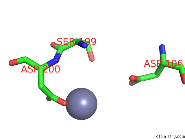
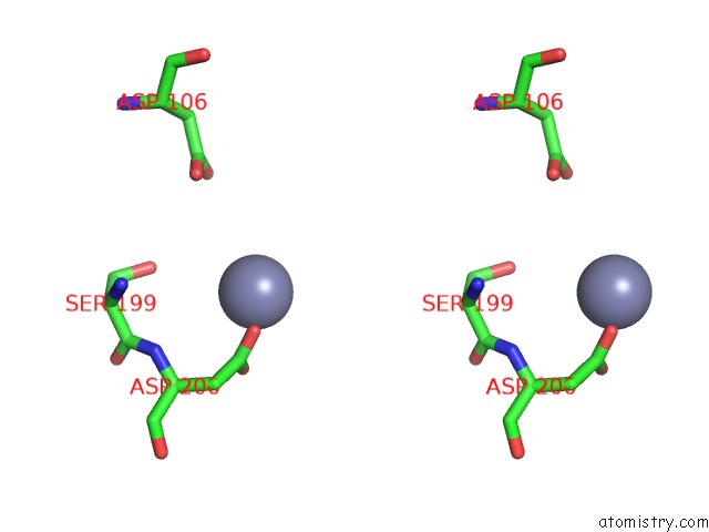
Zinc binding site 1 out of 3 in 1jaz
Go back to
Zinc binding site 1 out
of 3 in the Crystal Structure of Monoclinic Form of D90E Mutant of Escherichia Coli Asparaginase II
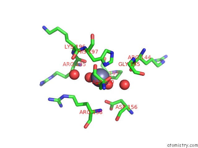
Mono view
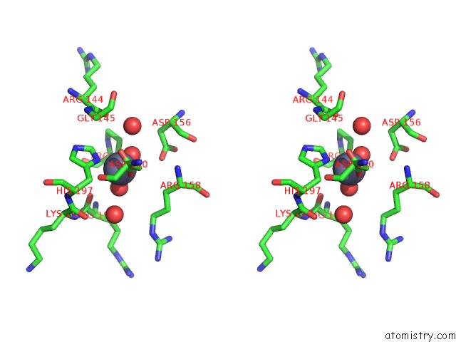
Stereo pair view
Mono view
Stereo pair view
A full contact list of Zinc with other atoms in the Zn binding
site number 1 of Crystal Structure of Monoclinic Form of D90E Mutant of Escherichia Coli Asparaginase II within 5.0Å range:
|
Zinc binding site 2 out of 3 in 1jaz
Go back to
Zinc binding site 2 out
of 3 in the Crystal Structure of Monoclinic Form of D90E Mutant of Escherichia Coli Asparaginase II
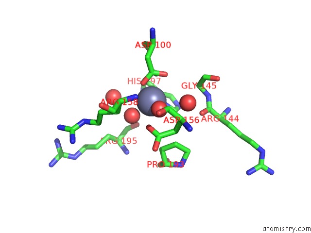
Mono view
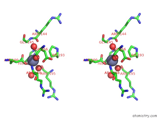
Stereo pair view
Mono view
Stereo pair view
A full contact list of Zinc with other atoms in the Zn binding
site number 2 of Crystal Structure of Monoclinic Form of D90E Mutant of Escherichia Coli Asparaginase II within 5.0Å range:
|
Zinc binding site 3 out of 3 in 1jaz
Go back to
Zinc binding site 3 out
of 3 in the Crystal Structure of Monoclinic Form of D90E Mutant of Escherichia Coli Asparaginase II
Mono view
Stereo pair view
Mono view
Stereo pair view
A full contact list of Zinc with other atoms in the Zn binding
site number 3 of Crystal Structure of Monoclinic Form of D90E Mutant of Escherichia Coli Asparaginase II within 5.0Å range:
|
Reference:
D.Borek,
M.Kozak,
J.Pei,
M.Jaskolski.
Crystal Structure of Active Site Mutant of Antileukemic L-Asparaginase Reveals Conserved Zinc-Binding Site. Febs J. V. 281 4097 2014.
ISSN: ISSN 1742-464X
PubMed: 25040257
DOI: 10.1111/FEBS.12906
Page generated: Sun Oct 13 03:28:24 2024
ISSN: ISSN 1742-464X
PubMed: 25040257
DOI: 10.1111/FEBS.12906
Last articles
Zn in 9J0NZn in 9J0O
Zn in 9J0P
Zn in 9FJX
Zn in 9EKB
Zn in 9C0F
Zn in 9CAH
Zn in 9CH0
Zn in 9CH3
Zn in 9CH1