Zinc »
PDB 1iml-1jan »
1j9y »
Zinc in PDB 1j9y: Crystal Structure of Mannanase 26A From Pseudomonas Cellulosa
Enzymatic activity of Crystal Structure of Mannanase 26A From Pseudomonas Cellulosa
All present enzymatic activity of Crystal Structure of Mannanase 26A From Pseudomonas Cellulosa:
3.2.1.78;
3.2.1.78;
Protein crystallography data
The structure of Crystal Structure of Mannanase 26A From Pseudomonas Cellulosa, PDB code: 1j9y
was solved by
D.Hogg,
E.-J.Woo,
D.N.Bolam,
V.A.Mckie,
H.J.Gilbert,
R.W.Pickersgill,
with X-Ray Crystallography technique. A brief refinement statistics is given in the table below:
| Resolution Low / High (Å) | 12.50 / 1.85 |
| Space group | P 41 |
| Cell size a, b, c (Å), α, β, γ (°) | 93.240, 93.240, 54.830, 90.00, 90.00, 90.00 |
| R / Rfree (%) | 18.2 / 20 |
Zinc Binding Sites:
The binding sites of Zinc atom in the Crystal Structure of Mannanase 26A From Pseudomonas Cellulosa
(pdb code 1j9y). This binding sites where shown within
5.0 Angstroms radius around Zinc atom.
In total 4 binding sites of Zinc where determined in the Crystal Structure of Mannanase 26A From Pseudomonas Cellulosa, PDB code: 1j9y:
Jump to Zinc binding site number: 1; 2; 3; 4;
In total 4 binding sites of Zinc where determined in the Crystal Structure of Mannanase 26A From Pseudomonas Cellulosa, PDB code: 1j9y:
Jump to Zinc binding site number: 1; 2; 3; 4;
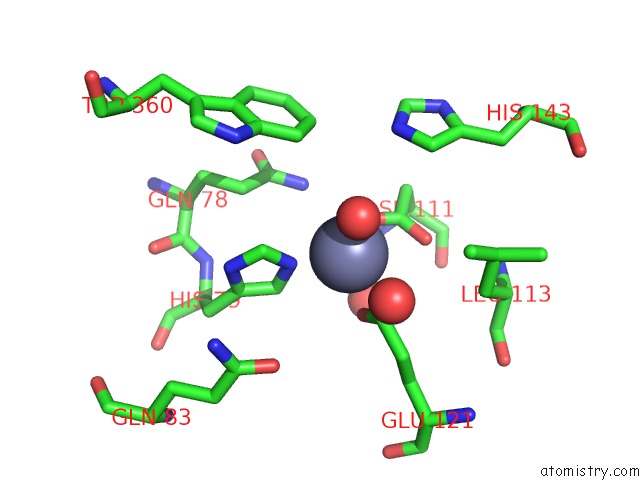
Zinc binding site 1 out of 4 in 1j9y
Go back to
Zinc binding site 1 out
of 4 in the Crystal Structure of Mannanase 26A From Pseudomonas Cellulosa
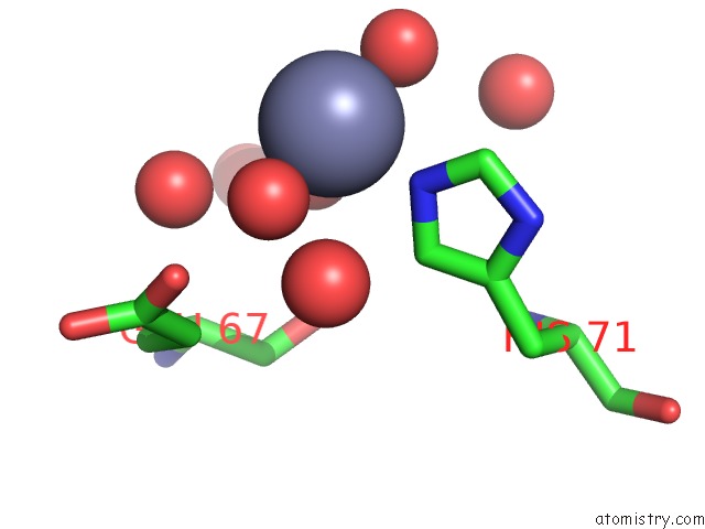
Mono view
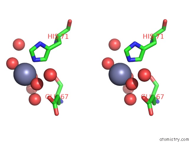
Stereo pair view
Mono view
Stereo pair view
A full contact list of Zinc with other atoms in the Zn binding
site number 1 of Crystal Structure of Mannanase 26A From Pseudomonas Cellulosa within 5.0Å range:
|
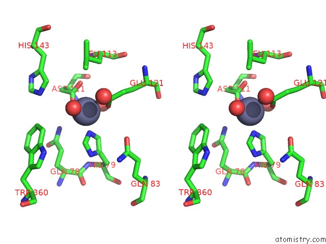
Zinc binding site 2 out of 4 in 1j9y
Go back to
Zinc binding site 2 out
of 4 in the Crystal Structure of Mannanase 26A From Pseudomonas Cellulosa
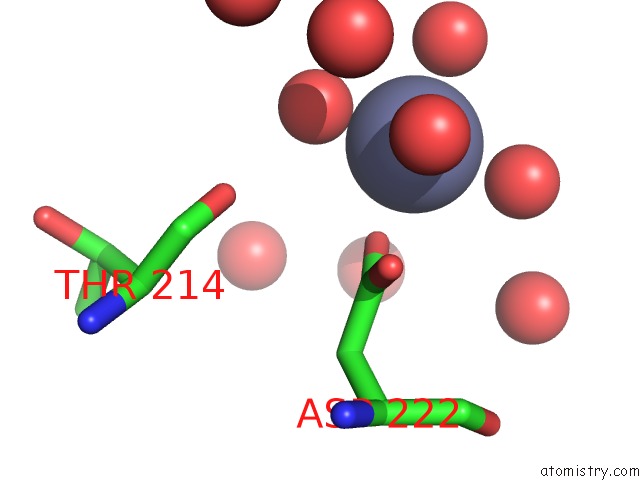
Mono view
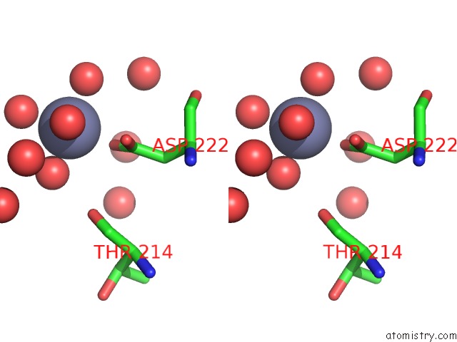
Stereo pair view
Mono view
Stereo pair view
A full contact list of Zinc with other atoms in the Zn binding
site number 2 of Crystal Structure of Mannanase 26A From Pseudomonas Cellulosa within 5.0Å range:
|
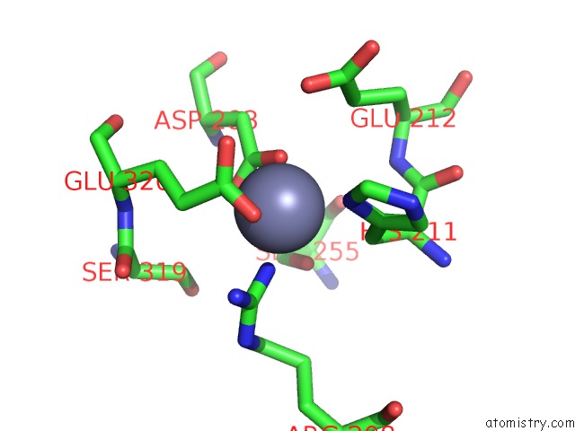
Zinc binding site 3 out of 4 in 1j9y
Go back to
Zinc binding site 3 out
of 4 in the Crystal Structure of Mannanase 26A From Pseudomonas Cellulosa
Mono view
Stereo pair view
Mono view
Stereo pair view
A full contact list of Zinc with other atoms in the Zn binding
site number 3 of Crystal Structure of Mannanase 26A From Pseudomonas Cellulosa within 5.0Å range:
|
Zinc binding site 4 out of 4 in 1j9y
Go back to
Zinc binding site 4 out
of 4 in the Crystal Structure of Mannanase 26A From Pseudomonas Cellulosa
Mono view
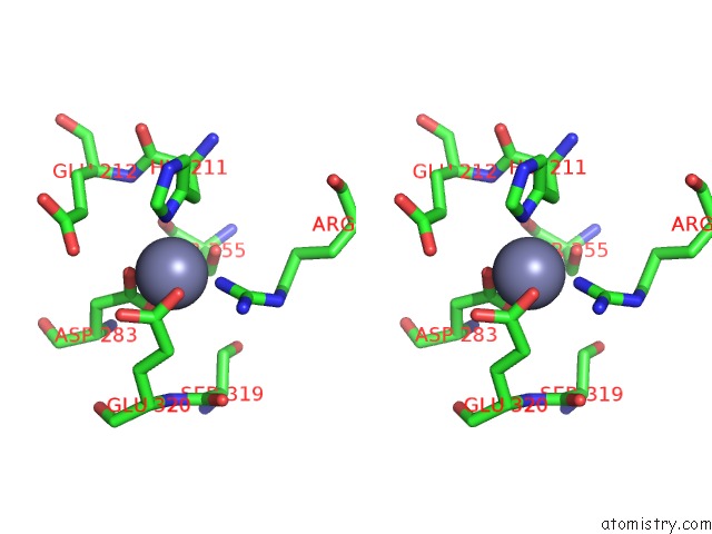
Stereo pair view
Mono view
Stereo pair view
A full contact list of Zinc with other atoms in the Zn binding
site number 4 of Crystal Structure of Mannanase 26A From Pseudomonas Cellulosa within 5.0Å range:
|
Reference:
D.Hogg,
E.-J.Woo,
D.N.Bolam,
V.A.Mckie,
H.J.Gilbert,
R.W.Pickersgill.
Crystal Structure of Mannanase 26A From Pseudomonas Cellulosa and Analysis of Residues Involved in Substrate Binding J.Biol.Chem. V. 276 31186 2001.
ISSN: ISSN 0021-9258
PubMed: 11382747
DOI: 10.1074/JBC.M010290200
Page generated: Sun Oct 13 03:27:06 2024
ISSN: ISSN 0021-9258
PubMed: 11382747
DOI: 10.1074/JBC.M010290200
Last articles
Zn in 9J0NZn in 9J0O
Zn in 9J0P
Zn in 9FJX
Zn in 9EKB
Zn in 9C0F
Zn in 9CAH
Zn in 9CH0
Zn in 9CH3
Zn in 9CH1