Zinc »
PDB 1iml-1jan »
1j9w »
Zinc in PDB 1j9w: Solution Structure of the Cai Michigan 1 Variant
Enzymatic activity of Solution Structure of the Cai Michigan 1 Variant
All present enzymatic activity of Solution Structure of the Cai Michigan 1 Variant:
4.2.1.1;
4.2.1.1;
Protein crystallography data
The structure of Solution Structure of the Cai Michigan 1 Variant, PDB code: 1j9w
was solved by
F.Briganti,
M.Ferraroni,
W.R.Chedwiggen,
A.Scozzafava,
C.T.Supuran,
S.Tilli,
with X-Ray Crystallography technique. A brief refinement statistics is given in the table below:
| Resolution Low / High (Å) | 15.00 / 2.60 |
| Space group | P 21 21 21 |
| Cell size a, b, c (Å), α, β, γ (°) | 61.875, 71.732, 120.392, 90.00, 90.00, 90.00 |
| R / Rfree (%) | 20.5 / 31.1 |
Zinc Binding Sites:
The binding sites of Zinc atom in the Solution Structure of the Cai Michigan 1 Variant
(pdb code 1j9w). This binding sites where shown within
5.0 Angstroms radius around Zinc atom.
In total 2 binding sites of Zinc where determined in the Solution Structure of the Cai Michigan 1 Variant, PDB code: 1j9w:
Jump to Zinc binding site number: 1; 2;
In total 2 binding sites of Zinc where determined in the Solution Structure of the Cai Michigan 1 Variant, PDB code: 1j9w:
Jump to Zinc binding site number: 1; 2;
Zinc binding site 1 out of 2 in 1j9w
Go back to
Zinc binding site 1 out
of 2 in the Solution Structure of the Cai Michigan 1 Variant
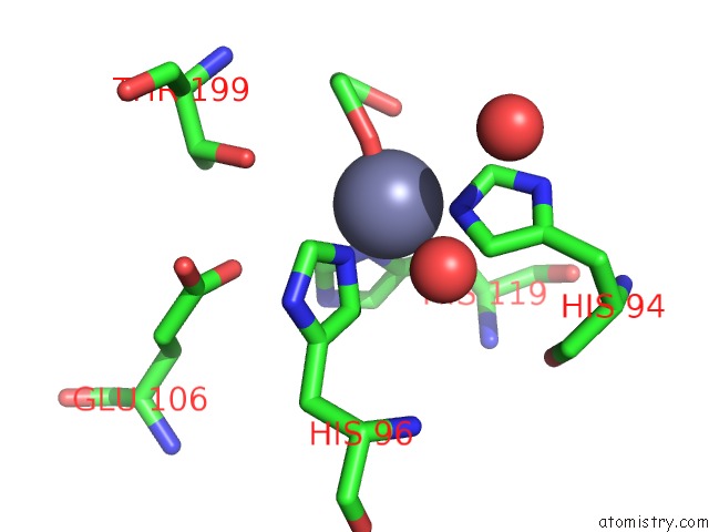
Mono view
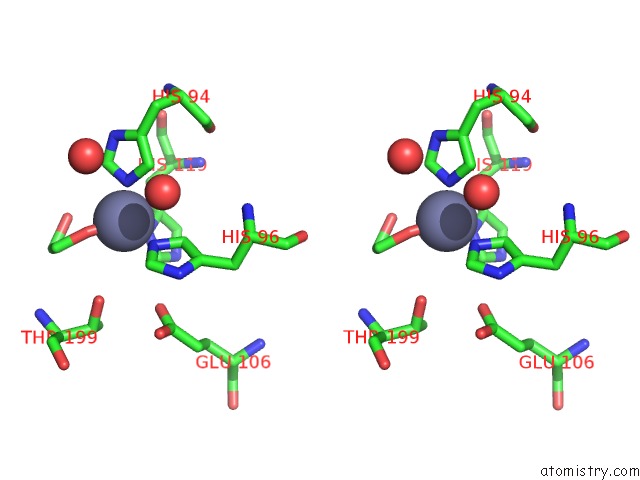
Stereo pair view
Mono view
Stereo pair view
A full contact list of Zinc with other atoms in the Zn binding
site number 1 of Solution Structure of the Cai Michigan 1 Variant within 5.0Å range:
|
Zinc binding site 2 out of 2 in 1j9w
Go back to
Zinc binding site 2 out
of 2 in the Solution Structure of the Cai Michigan 1 Variant
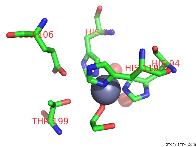
Mono view
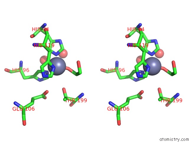
Stereo pair view
Mono view
Stereo pair view
A full contact list of Zinc with other atoms in the Zn binding
site number 2 of Solution Structure of the Cai Michigan 1 Variant within 5.0Å range:
|
Reference:
M.Ferraroni,
S.Tilli,
F.Briganti,
W.R.Chegwidden,
C.T.Supuran,
K.E.Wiebauer,
R.E.Tashian,
A.Scozzafava.
Crystal Structure of A Zinc-Activated Variant of Human Carbonic Anhydrase I, Ca I Michigan 1: Evidence For A Second Zinc Binding Site Involving Arginine Coordination. Biochemistry V. 41 6237 2002.
ISSN: ISSN 0006-2960
PubMed: 12009884
DOI: 10.1021/BI0120446
Page generated: Sun Oct 13 03:26:21 2024
ISSN: ISSN 0006-2960
PubMed: 12009884
DOI: 10.1021/BI0120446
Last articles
Zn in 9J0NZn in 9J0O
Zn in 9J0P
Zn in 9FJX
Zn in 9EKB
Zn in 9C0F
Zn in 9CAH
Zn in 9CH0
Zn in 9CH3
Zn in 9CH1