Zinc »
PDB 1iml-1jan »
1j6w »
Zinc in PDB 1j6w: Crystal Structure of Haemophilus Influenzae Luxs
Protein crystallography data
The structure of Crystal Structure of Haemophilus Influenzae Luxs, PDB code: 1j6w
was solved by
H.A.Lewis,
E.B.Furlong,
M.G.Bergseid,
W.E.Sanderson,
S.G.Buchanan,
with X-Ray Crystallography technique. A brief refinement statistics is given in the table below:
| Resolution Low / High (Å) | 30.00 / 2.10 |
| Space group | P 42 21 2 |
| Cell size a, b, c (Å), α, β, γ (°) | 129.669, 129.669, 53.770, 90.00, 90.00, 90.00 |
| R / Rfree (%) | 21.3 / 23.8 |
Zinc Binding Sites:
The binding sites of Zinc atom in the Crystal Structure of Haemophilus Influenzae Luxs
(pdb code 1j6w). This binding sites where shown within
5.0 Angstroms radius around Zinc atom.
In total 2 binding sites of Zinc where determined in the Crystal Structure of Haemophilus Influenzae Luxs, PDB code: 1j6w:
Jump to Zinc binding site number: 1; 2;
In total 2 binding sites of Zinc where determined in the Crystal Structure of Haemophilus Influenzae Luxs, PDB code: 1j6w:
Jump to Zinc binding site number: 1; 2;
Zinc binding site 1 out of 2 in 1j6w
Go back to
Zinc binding site 1 out
of 2 in the Crystal Structure of Haemophilus Influenzae Luxs
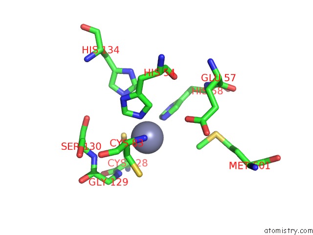
Mono view
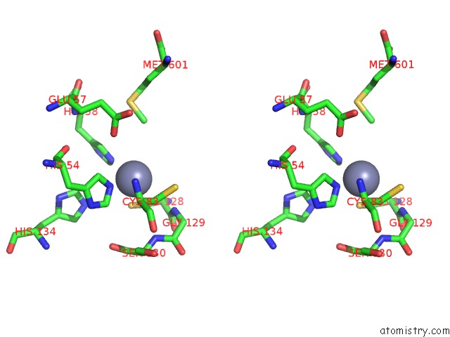
Stereo pair view
Mono view
Stereo pair view
A full contact list of Zinc with other atoms in the Zn binding
site number 1 of Crystal Structure of Haemophilus Influenzae Luxs within 5.0Å range:
|
Zinc binding site 2 out of 2 in 1j6w
Go back to
Zinc binding site 2 out
of 2 in the Crystal Structure of Haemophilus Influenzae Luxs
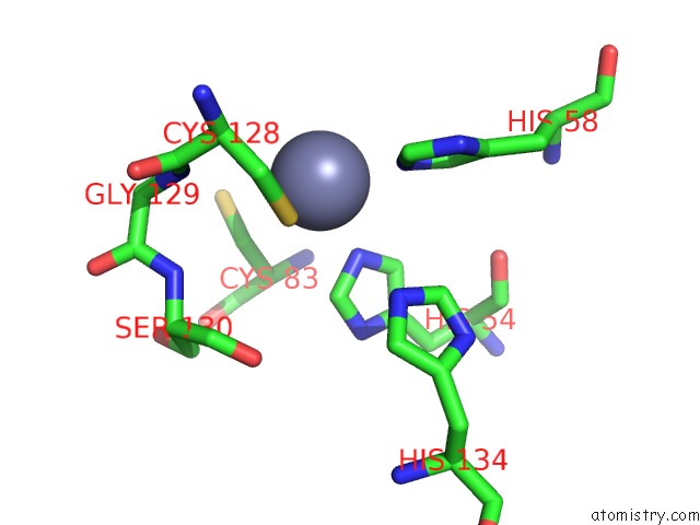
Mono view
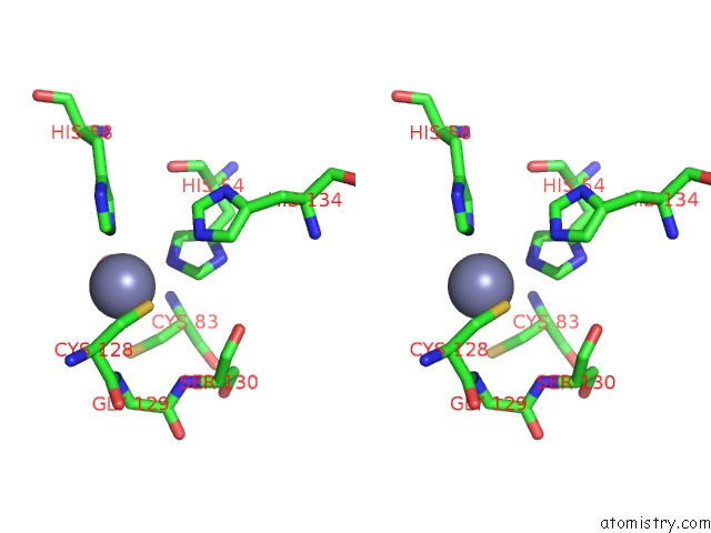
Stereo pair view
Mono view
Stereo pair view
A full contact list of Zinc with other atoms in the Zn binding
site number 2 of Crystal Structure of Haemophilus Influenzae Luxs within 5.0Å range:
|
Reference:
H.A.Lewis,
E.B.Furlong,
B.Laubert,
G.A.Eroshkina,
Y.Batiyenko,
J.M.Adams,
M.G.Bergseid,
C.D.Marsh,
T.S.Peat,
W.E.Sanderson,
J.M.Sauder,
S.G.Buchanan.
A Structural Genomics Approach to the Study of Quorum Sensing: Crystal Structures of Three Luxs Orthologs. Structure V. 9 527 2001.
ISSN: ISSN 0969-2126
PubMed: 11435117
DOI: 10.1016/S0969-2126(01)00613-X
Page generated: Sun Oct 13 03:22:46 2024
ISSN: ISSN 0969-2126
PubMed: 11435117
DOI: 10.1016/S0969-2126(01)00613-X
Last articles
Zn in 9J0NZn in 9J0O
Zn in 9J0P
Zn in 9FJX
Zn in 9EKB
Zn in 9C0F
Zn in 9CAH
Zn in 9CH0
Zn in 9CH3
Zn in 9CH1