Zinc »
PDB 1iml-1jan »
1itu »
Zinc in PDB 1itu: Human Renal Dipeptidase Complexed with Cilastatin
Enzymatic activity of Human Renal Dipeptidase Complexed with Cilastatin
All present enzymatic activity of Human Renal Dipeptidase Complexed with Cilastatin:
3.4.13.19;
3.4.13.19;
Protein crystallography data
The structure of Human Renal Dipeptidase Complexed with Cilastatin, PDB code: 1itu
was solved by
Y.Nitanai,
Y.Satow,
H.Adachi,
M.Tsujimoto,
with X-Ray Crystallography technique. A brief refinement statistics is given in the table below:
| Resolution Low / High (Å) | 10.00 / 2.00 |
| Space group | P 1 21 1 |
| Cell size a, b, c (Å), α, β, γ (°) | 80.179, 79.491, 56.951, 90.00, 96.34, 90.00 |
| R / Rfree (%) | 18.5 / 25.4 |
Zinc Binding Sites:
The binding sites of Zinc atom in the Human Renal Dipeptidase Complexed with Cilastatin
(pdb code 1itu). This binding sites where shown within
5.0 Angstroms radius around Zinc atom.
In total 4 binding sites of Zinc where determined in the Human Renal Dipeptidase Complexed with Cilastatin, PDB code: 1itu:
Jump to Zinc binding site number: 1; 2; 3; 4;
In total 4 binding sites of Zinc where determined in the Human Renal Dipeptidase Complexed with Cilastatin, PDB code: 1itu:
Jump to Zinc binding site number: 1; 2; 3; 4;
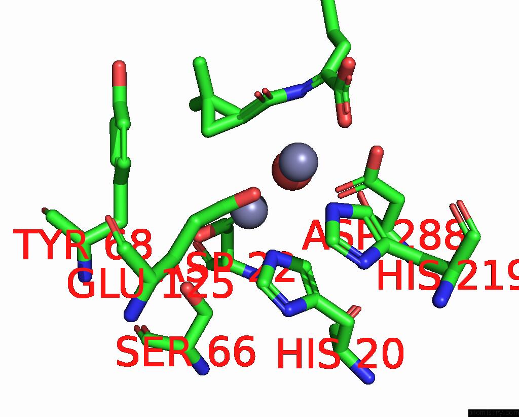
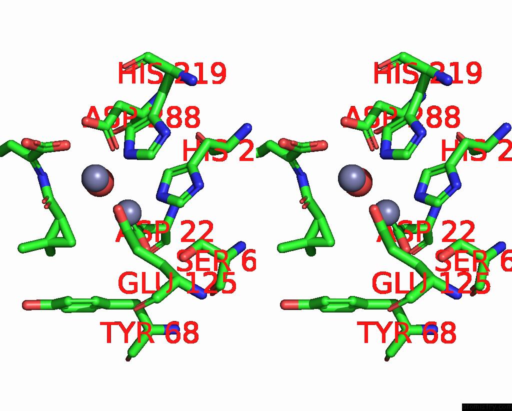
Zinc binding site 1 out of 4 in 1itu
Go back to
Zinc binding site 1 out
of 4 in the Human Renal Dipeptidase Complexed with Cilastatin
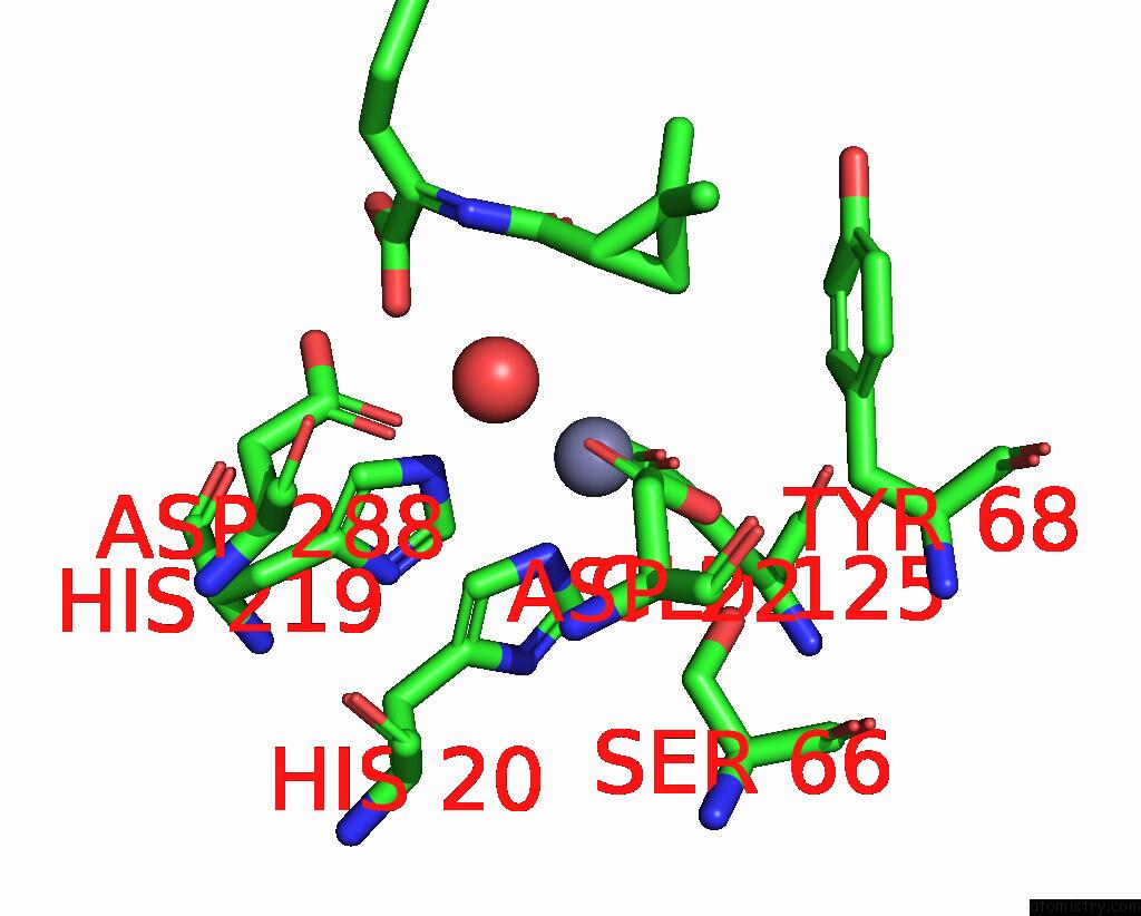
Mono view
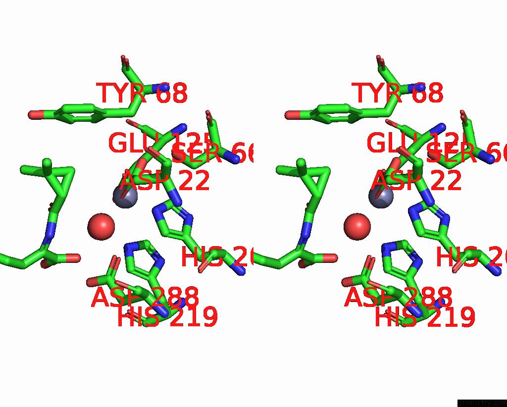
Stereo pair view
Mono view
Stereo pair view
A full contact list of Zinc with other atoms in the Zn binding
site number 1 of Human Renal Dipeptidase Complexed with Cilastatin within 5.0Å range:
|
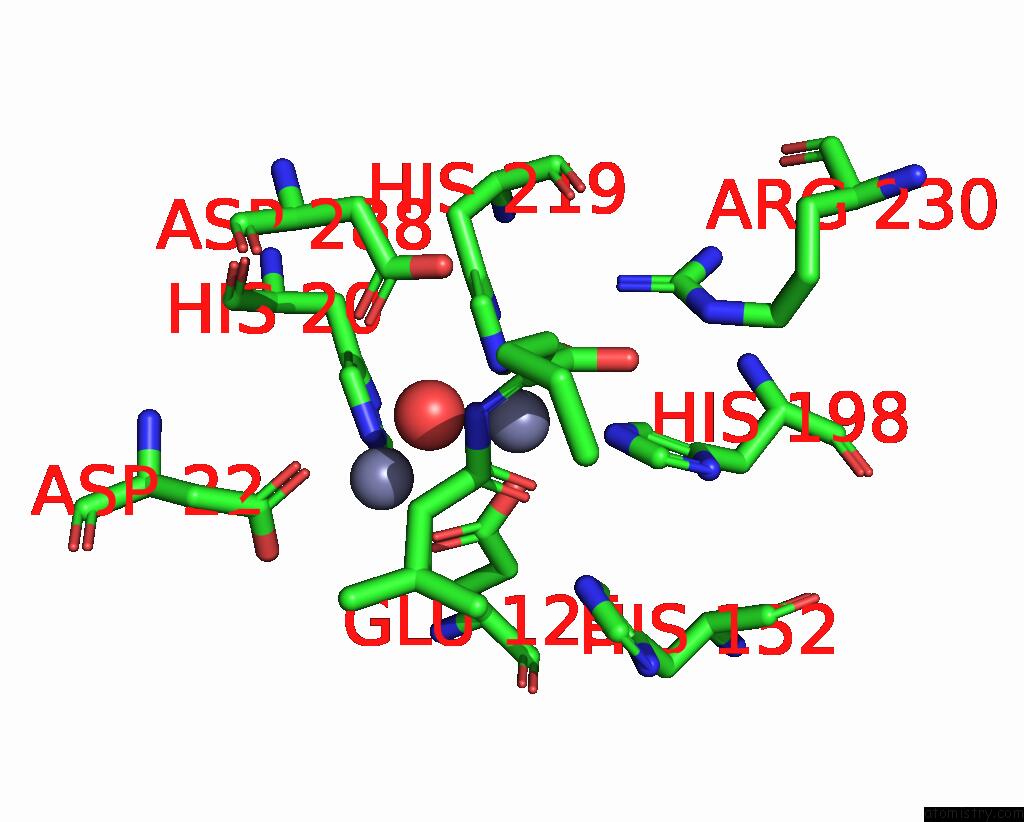
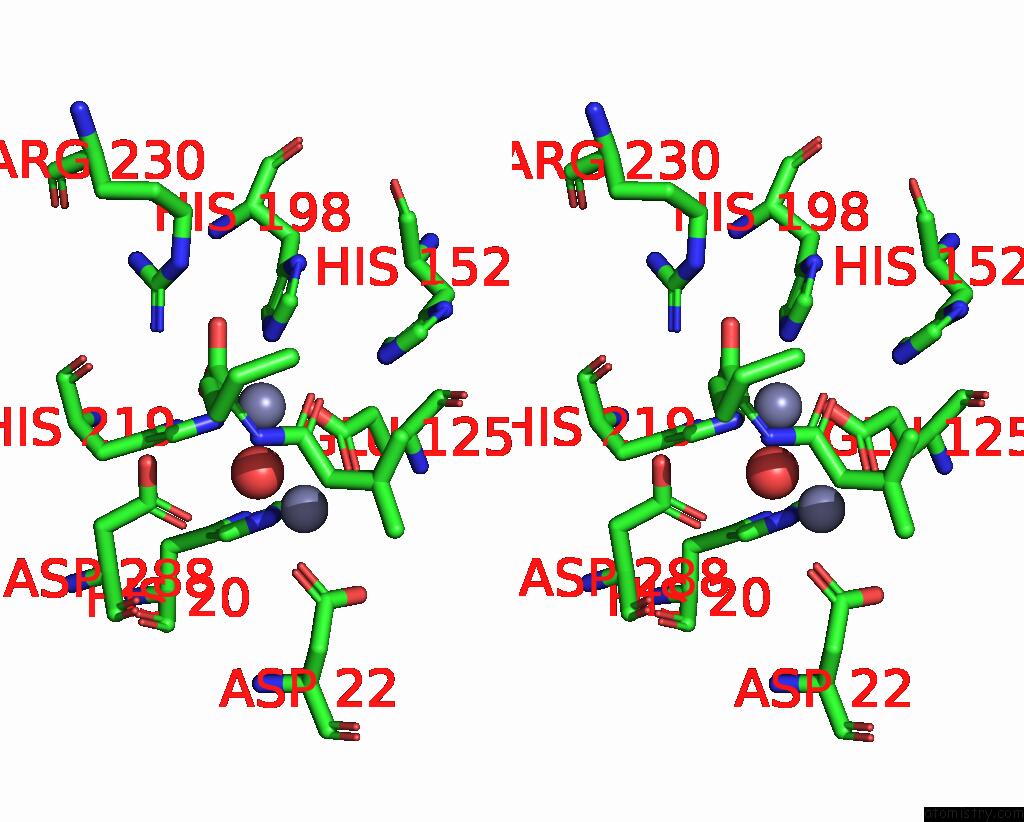
Zinc binding site 2 out of 4 in 1itu
Go back to
Zinc binding site 2 out
of 4 in the Human Renal Dipeptidase Complexed with Cilastatin
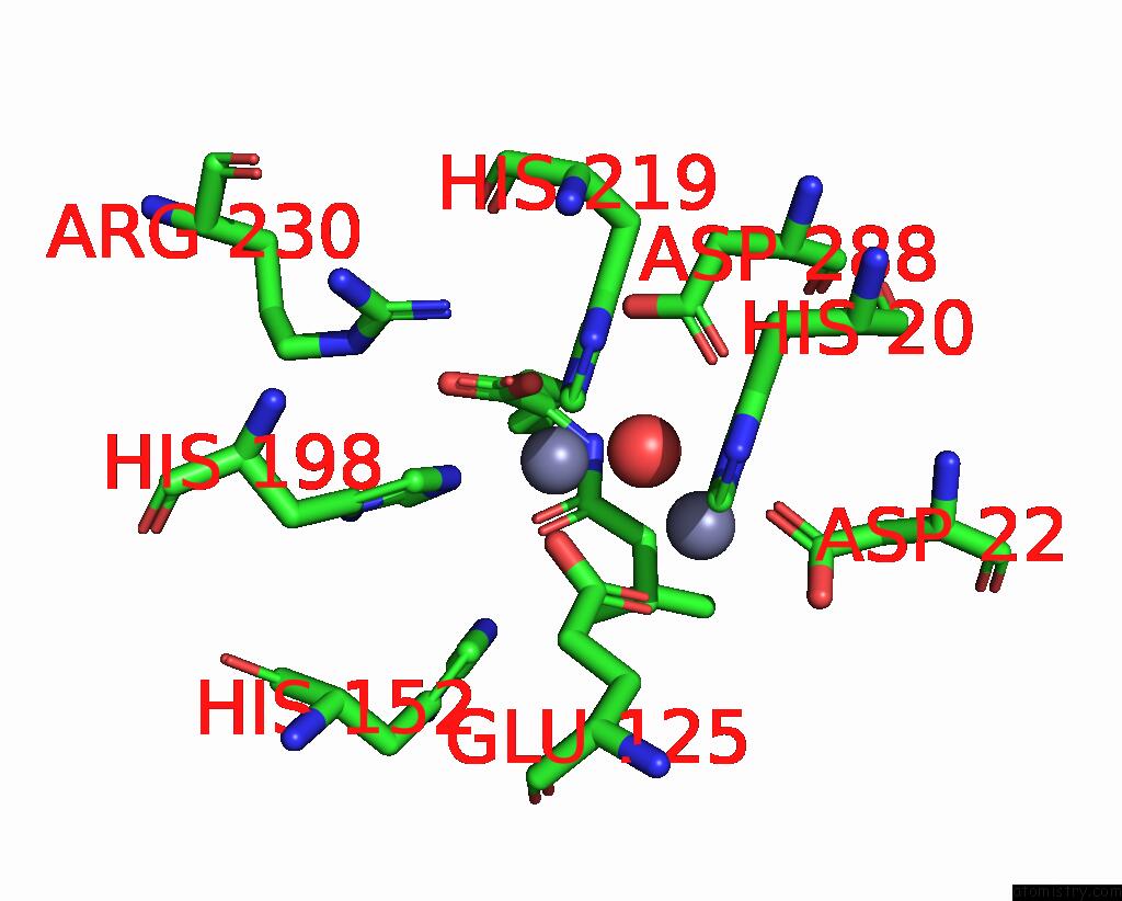
Mono view
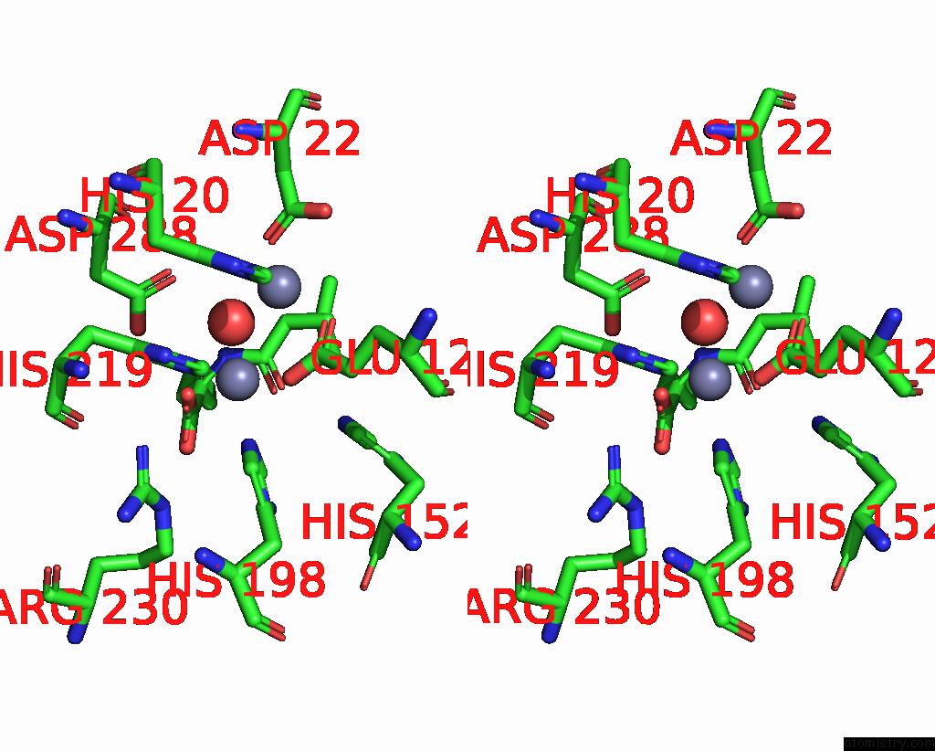
Stereo pair view
Mono view
Stereo pair view
A full contact list of Zinc with other atoms in the Zn binding
site number 2 of Human Renal Dipeptidase Complexed with Cilastatin within 5.0Å range:
|
Zinc binding site 3 out of 4 in 1itu
Go back to
Zinc binding site 3 out
of 4 in the Human Renal Dipeptidase Complexed with Cilastatin
Mono view
Stereo pair view
Mono view
Stereo pair view
A full contact list of Zinc with other atoms in the Zn binding
site number 3 of Human Renal Dipeptidase Complexed with Cilastatin within 5.0Å range:
|
Zinc binding site 4 out of 4 in 1itu
Go back to
Zinc binding site 4 out
of 4 in the Human Renal Dipeptidase Complexed with Cilastatin
Mono view
Stereo pair view
Mono view
Stereo pair view
A full contact list of Zinc with other atoms in the Zn binding
site number 4 of Human Renal Dipeptidase Complexed with Cilastatin within 5.0Å range:
|
Reference:
Y.Nitanai,
Y.Satow,
H.Adachi,
M.Tsujimoto.
Crystal Structure of Human Renal Dipeptidase Involved in Beta-Lactam Hydrolysis J.Mol.Biol. V. 321 177 2002.
ISSN: ISSN 0022-2836
PubMed: 12144777
DOI: 10.1016/S0022-2836(02)00632-0
Page generated: Sun Oct 13 03:13:40 2024
ISSN: ISSN 0022-2836
PubMed: 12144777
DOI: 10.1016/S0022-2836(02)00632-0
Last articles
Zn in 9J0NZn in 9J0O
Zn in 9J0P
Zn in 9FJX
Zn in 9EKB
Zn in 9C0F
Zn in 9CAH
Zn in 9CH0
Zn in 9CH3
Zn in 9CH1