Zinc »
PDB 1iml-1jan »
1iq8 »
Zinc in PDB 1iq8: Crystal Structure of Archaeosine Trna-Guanine Transglycosylase From Pyrococcus Horikoshii
Enzymatic activity of Crystal Structure of Archaeosine Trna-Guanine Transglycosylase From Pyrococcus Horikoshii
All present enzymatic activity of Crystal Structure of Archaeosine Trna-Guanine Transglycosylase From Pyrococcus Horikoshii:
2.4.2.29;
2.4.2.29;
Protein crystallography data
The structure of Crystal Structure of Archaeosine Trna-Guanine Transglycosylase From Pyrococcus Horikoshii, PDB code: 1iq8
was solved by
R.Ishitani,
O.Nureki,
S.Fukai,
T.Kijimoto,
N.Nameki,
M.Watanabe,
H.Kondo,
M.Sekine,
N.Okada,
S.Nishimura,
S.Yokoyama,
Riken Structuralgenomics/Proteomics Initiative (Rsgi),
with X-Ray Crystallography technique. A brief refinement statistics is given in the table below:
| Resolution Low / High (Å) | 50.00 / 2.20 |
| Space group | P 43 21 2 |
| Cell size a, b, c (Å), α, β, γ (°) | 99.282, 99.282, 363.740, 90.00, 90.00, 90.00 |
| R / Rfree (%) | 22.7 / 26.1 |
Other elements in 1iq8:
The structure of Crystal Structure of Archaeosine Trna-Guanine Transglycosylase From Pyrococcus Horikoshii also contains other interesting chemical elements:
| Magnesium | (Mg) | 2 atoms |
Zinc Binding Sites:
The binding sites of Zinc atom in the Crystal Structure of Archaeosine Trna-Guanine Transglycosylase From Pyrococcus Horikoshii
(pdb code 1iq8). This binding sites where shown within
5.0 Angstroms radius around Zinc atom.
In total 2 binding sites of Zinc where determined in the Crystal Structure of Archaeosine Trna-Guanine Transglycosylase From Pyrococcus Horikoshii, PDB code: 1iq8:
Jump to Zinc binding site number: 1; 2;
In total 2 binding sites of Zinc where determined in the Crystal Structure of Archaeosine Trna-Guanine Transglycosylase From Pyrococcus Horikoshii, PDB code: 1iq8:
Jump to Zinc binding site number: 1; 2;
Zinc binding site 1 out of 2 in 1iq8
Go back to
Zinc binding site 1 out
of 2 in the Crystal Structure of Archaeosine Trna-Guanine Transglycosylase From Pyrococcus Horikoshii
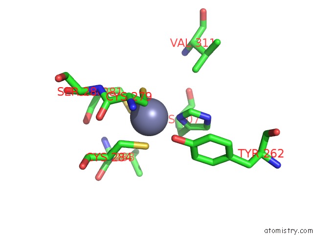
Mono view
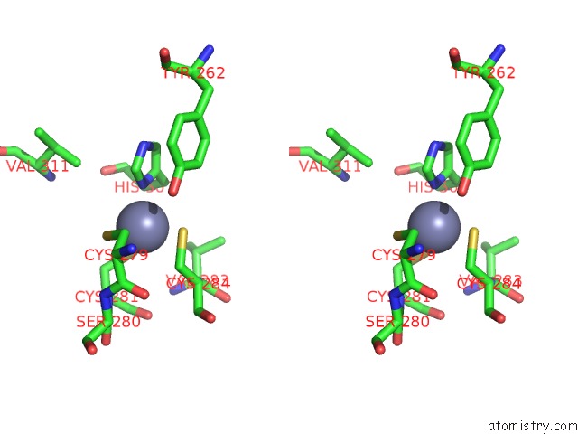
Stereo pair view
Mono view
Stereo pair view
A full contact list of Zinc with other atoms in the Zn binding
site number 1 of Crystal Structure of Archaeosine Trna-Guanine Transglycosylase From Pyrococcus Horikoshii within 5.0Å range:
|
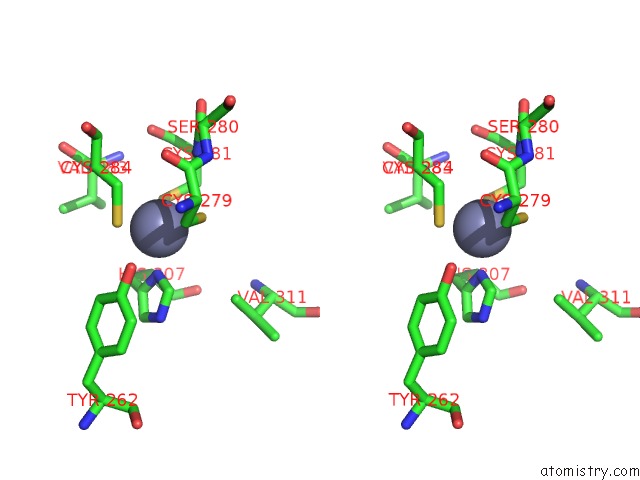
Zinc binding site 2 out of 2 in 1iq8
Go back to
Zinc binding site 2 out
of 2 in the Crystal Structure of Archaeosine Trna-Guanine Transglycosylase From Pyrococcus Horikoshii
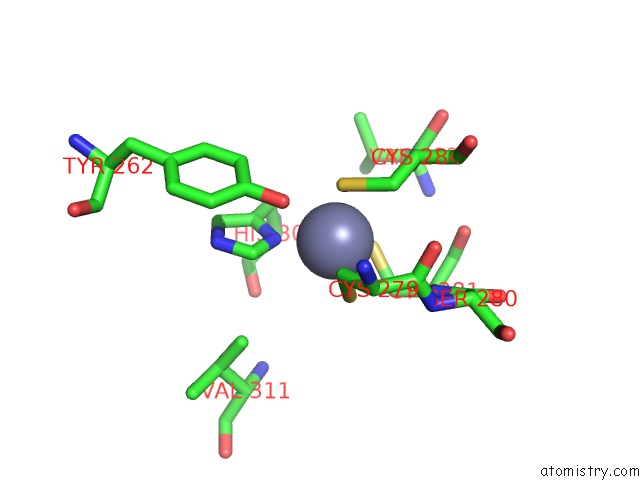
Mono view
Stereo pair view
Mono view
Stereo pair view
A full contact list of Zinc with other atoms in the Zn binding
site number 2 of Crystal Structure of Archaeosine Trna-Guanine Transglycosylase From Pyrococcus Horikoshii within 5.0Å range:
|
Reference:
R.Ishitani,
O.Nureki,
S.Fukai,
T.Kijimoto,
N.Nameki,
M.Watanabe,
H.Kondo,
M.Sekine,
N.Okada,
S.Nishimura,
S.Yokoyama.
Crystal Structure of Archaeosine Trna-Guanine Transglycosylase. J.Mol.Biol. V. 318 665 2002.
ISSN: ISSN 0022-2836
PubMed: 12054814
DOI: 10.1016/S0022-2836(02)00090-6
Page generated: Sun Oct 13 03:09:22 2024
ISSN: ISSN 0022-2836
PubMed: 12054814
DOI: 10.1016/S0022-2836(02)00090-6
Last articles
Zn in 9J0NZn in 9J0O
Zn in 9J0P
Zn in 9FJX
Zn in 9EKB
Zn in 9C0F
Zn in 9CAH
Zn in 9CH0
Zn in 9CH3
Zn in 9CH1