Zinc »
PDB 1i96-1im5 »
1ici »
Zinc in PDB 1ici: Crystal Structure of A SIR2 Homolog-Nad Complex
Protein crystallography data
The structure of Crystal Structure of A SIR2 Homolog-Nad Complex, PDB code: 1ici
was solved by
J.Min,
J.Landry,
R.Sternglanz,
R.-M.Xu,
with X-Ray Crystallography technique. A brief refinement statistics is given in the table below:
| Resolution Low / High (Å) | 40.00 / 2.10 |
| Space group | C 1 2 1 |
| Cell size a, b, c (Å), α, β, γ (°) | 65.410, 94.540, 93.780, 90.00, 95.29, 90.00 |
| R / Rfree (%) | 19.9 / 24.9 |
Zinc Binding Sites:
The binding sites of Zinc atom in the Crystal Structure of A SIR2 Homolog-Nad Complex
(pdb code 1ici). This binding sites where shown within
5.0 Angstroms radius around Zinc atom.
In total 2 binding sites of Zinc where determined in the Crystal Structure of A SIR2 Homolog-Nad Complex, PDB code: 1ici:
Jump to Zinc binding site number: 1; 2;
In total 2 binding sites of Zinc where determined in the Crystal Structure of A SIR2 Homolog-Nad Complex, PDB code: 1ici:
Jump to Zinc binding site number: 1; 2;
Zinc binding site 1 out of 2 in 1ici
Go back to
Zinc binding site 1 out
of 2 in the Crystal Structure of A SIR2 Homolog-Nad Complex
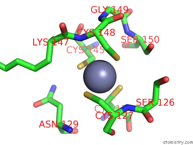
Mono view
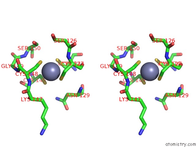
Stereo pair view
Mono view
Stereo pair view
A full contact list of Zinc with other atoms in the Zn binding
site number 1 of Crystal Structure of A SIR2 Homolog-Nad Complex within 5.0Å range:
|
Zinc binding site 2 out of 2 in 1ici
Go back to
Zinc binding site 2 out
of 2 in the Crystal Structure of A SIR2 Homolog-Nad Complex
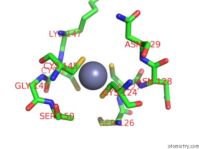
Mono view
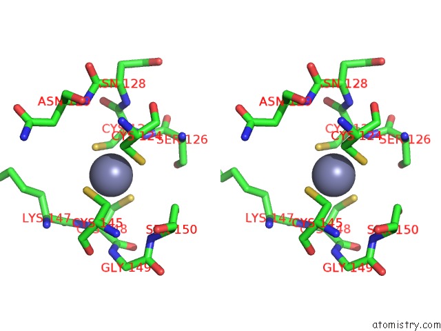
Stereo pair view
Mono view
Stereo pair view
A full contact list of Zinc with other atoms in the Zn binding
site number 2 of Crystal Structure of A SIR2 Homolog-Nad Complex within 5.0Å range:
|
Reference:
J.Min,
J.Landry,
R.Sternglanz,
R.M.Xu.
Crystal Structure of A SIR2 Homolog-Nad Complex. Cell(Cambridge,Mass.) V. 105 269 2001.
ISSN: ISSN 0092-8674
PubMed: 11336676
DOI: 10.1016/S0092-8674(01)00317-8
Page generated: Sun Oct 13 03:05:05 2024
ISSN: ISSN 0092-8674
PubMed: 11336676
DOI: 10.1016/S0092-8674(01)00317-8
Last articles
Zn in 9J0NZn in 9J0O
Zn in 9J0P
Zn in 9FJX
Zn in 9EKB
Zn in 9C0F
Zn in 9CAH
Zn in 9CH0
Zn in 9CH3
Zn in 9CH1