Zinc »
PDB 1i96-1im5 »
1ia7 »
Zinc in PDB 1ia7: Crystal Structure of the Cellulase CEL9M of C. Cellulolyticium in Complex with Cellobiose
Enzymatic activity of Crystal Structure of the Cellulase CEL9M of C. Cellulolyticium in Complex with Cellobiose
All present enzymatic activity of Crystal Structure of the Cellulase CEL9M of C. Cellulolyticium in Complex with Cellobiose:
3.2.1.4;
3.2.1.4;
Protein crystallography data
The structure of Crystal Structure of the Cellulase CEL9M of C. Cellulolyticium in Complex with Cellobiose, PDB code: 1ia7
was solved by
G.Parsiegla,
A.Belaich,
J.P.Belaich,
R.Haser,
with X-Ray Crystallography technique. A brief refinement statistics is given in the table below:
| Resolution Low / High (Å) | 16.23 / 2.00 |
| Space group | P 1 21 1 |
| Cell size a, b, c (Å), α, β, γ (°) | 51.850, 52.380, 71.970, 90.00, 108.46, 90.00 |
| R / Rfree (%) | 16.8 / 22 |
Other elements in 1ia7:
The structure of Crystal Structure of the Cellulase CEL9M of C. Cellulolyticium in Complex with Cellobiose also contains other interesting chemical elements:
| Nickel | (Ni) | 1 atom |
| Calcium | (Ca) | 1 atom |
Zinc Binding Sites:
The binding sites of Zinc atom in the Crystal Structure of the Cellulase CEL9M of C. Cellulolyticium in Complex with Cellobiose
(pdb code 1ia7). This binding sites where shown within
5.0 Angstroms radius around Zinc atom.
In total only one binding site of Zinc was determined in the Crystal Structure of the Cellulase CEL9M of C. Cellulolyticium in Complex with Cellobiose, PDB code: 1ia7:
In total only one binding site of Zinc was determined in the Crystal Structure of the Cellulase CEL9M of C. Cellulolyticium in Complex with Cellobiose, PDB code: 1ia7:
Zinc binding site 1 out of 1 in 1ia7
Go back to
Zinc binding site 1 out
of 1 in the Crystal Structure of the Cellulase CEL9M of C. Cellulolyticium in Complex with Cellobiose
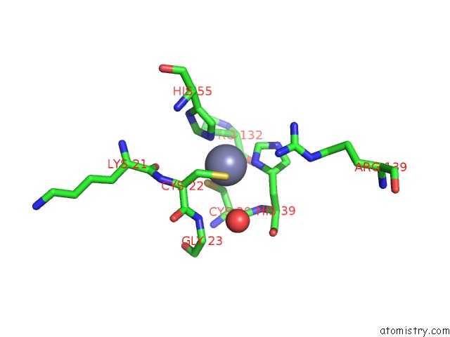
Mono view
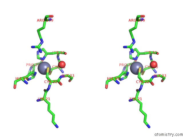
Stereo pair view
Mono view
Stereo pair view
A full contact list of Zinc with other atoms in the Zn binding
site number 1 of Crystal Structure of the Cellulase CEL9M of C. Cellulolyticium in Complex with Cellobiose within 5.0Å range:
|
Reference:
G.Parsiegla,
A.Belaich,
J.P.Belaich,
R.Haser.
Crystal Structure of the Cellulase CEL9M Enlightens Structure/Function Relationships of the Variable Catalytic Modules in Glycoside Hydrolases. Biochemistry V. 41 11134 2002.
ISSN: ISSN 0006-2960
PubMed: 12220178
DOI: 10.1021/BI025816M
Page generated: Sun Oct 13 03:00:34 2024
ISSN: ISSN 0006-2960
PubMed: 12220178
DOI: 10.1021/BI025816M
Last articles
Zn in 9J0NZn in 9J0O
Zn in 9J0P
Zn in 9FJX
Zn in 9EKB
Zn in 9C0F
Zn in 9CAH
Zn in 9CH0
Zn in 9CH3
Zn in 9CH1