Zinc »
PDB 1hxy-1i95 »
1i1i »
Zinc in PDB 1i1i: Neurolysin (Endopeptidase 24.16) Crystal Structure
Enzymatic activity of Neurolysin (Endopeptidase 24.16) Crystal Structure
All present enzymatic activity of Neurolysin (Endopeptidase 24.16) Crystal Structure:
3.4.24.16;
3.4.24.16;
Protein crystallography data
The structure of Neurolysin (Endopeptidase 24.16) Crystal Structure, PDB code: 1i1i
was solved by
C.K.Brown,
K.Madauss,
W.Lian,
W.D.Tolbert,
M.R.Beck,
D.W.Rodgers,
with X-Ray Crystallography technique. A brief refinement statistics is given in the table below:
| Resolution Low / High (Å) | 20.00 / 2.30 |
| Space group | P 21 21 2 |
| Cell size a, b, c (Å), α, β, γ (°) | 157.600, 87.700, 58.300, 90.00, 90.00, 90.00 |
| R / Rfree (%) | 22.4 / 26.8 |
Zinc Binding Sites:
The binding sites of Zinc atom in the Neurolysin (Endopeptidase 24.16) Crystal Structure
(pdb code 1i1i). This binding sites where shown within
5.0 Angstroms radius around Zinc atom.
In total 2 binding sites of Zinc where determined in the Neurolysin (Endopeptidase 24.16) Crystal Structure, PDB code: 1i1i:
Jump to Zinc binding site number: 1; 2;
In total 2 binding sites of Zinc where determined in the Neurolysin (Endopeptidase 24.16) Crystal Structure, PDB code: 1i1i:
Jump to Zinc binding site number: 1; 2;
Zinc binding site 1 out of 2 in 1i1i
Go back to
Zinc binding site 1 out
of 2 in the Neurolysin (Endopeptidase 24.16) Crystal Structure
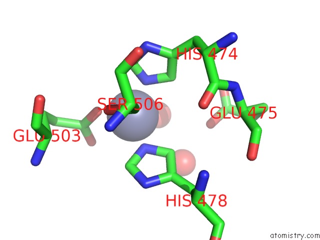
Mono view
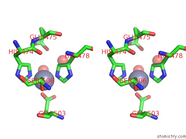
Stereo pair view
Mono view
Stereo pair view
A full contact list of Zinc with other atoms in the Zn binding
site number 1 of Neurolysin (Endopeptidase 24.16) Crystal Structure within 5.0Å range:
|
Zinc binding site 2 out of 2 in 1i1i
Go back to
Zinc binding site 2 out
of 2 in the Neurolysin (Endopeptidase 24.16) Crystal Structure
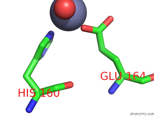
Mono view
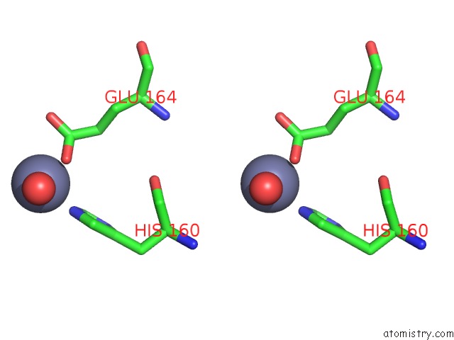
Stereo pair view
Mono view
Stereo pair view
A full contact list of Zinc with other atoms in the Zn binding
site number 2 of Neurolysin (Endopeptidase 24.16) Crystal Structure within 5.0Å range:
|
Reference:
C.K.Brown,
K.Madauss,
W.Lian,
M.R.Beck,
W.D.Tolbert,
D.W.Rodgers.
Structure of Neurolysin Reveals A Deep Channel That Limits Substrate Access. Proc.Natl.Acad.Sci.Usa V. 98 3127 2001.
ISSN: ISSN 0027-8424
PubMed: 11248043
DOI: 10.1073/PNAS.051633198
Page generated: Sun Oct 13 02:46:09 2024
ISSN: ISSN 0027-8424
PubMed: 11248043
DOI: 10.1073/PNAS.051633198
Last articles
Zn in 9J0NZn in 9J0O
Zn in 9J0P
Zn in 9FJX
Zn in 9EKB
Zn in 9C0F
Zn in 9CAH
Zn in 9CH0
Zn in 9CH3
Zn in 9CH1