Zinc »
PDB 1hlk-1hxr »
1hv5 »
Zinc in PDB 1hv5: Crystal Structure of the Stromelysin-3 (Mmp-11) Catalytic Domain Complexed with A Phosphinic Inhibitor
Protein crystallography data
The structure of Crystal Structure of the Stromelysin-3 (Mmp-11) Catalytic Domain Complexed with A Phosphinic Inhibitor, PDB code: 1hv5
was solved by
A.L.Gall,
M.Ruff,
R.Kannan,
P.Cuniasse,
A.Yiotakis,
V.Dive,
M.C.Rio,
P.Basset,
D.Moras,
with X-Ray Crystallography technique. A brief refinement statistics is given in the table below:
| Resolution Low / High (Å) | 19.89 / 2.60 |
| Space group | P 21 21 21 |
| Cell size a, b, c (Å), α, β, γ (°) | 140.100, 148.500, 91.400, 90.00, 90.00, 90.00 |
| R / Rfree (%) | 21.8 / 26.2 |
Other elements in 1hv5:
The structure of Crystal Structure of the Stromelysin-3 (Mmp-11) Catalytic Domain Complexed with A Phosphinic Inhibitor also contains other interesting chemical elements:
| Calcium | (Ca) | 6 atoms |
Zinc Binding Sites:
Pages:
>>> Page 1 <<< Page 2, Binding sites: 11 - 12;Binding sites:
The binding sites of Zinc atom in the Crystal Structure of the Stromelysin-3 (Mmp-11) Catalytic Domain Complexed with A Phosphinic Inhibitor (pdb code 1hv5). This binding sites where shown within 5.0 Angstroms radius around Zinc atom.In total 12 binding sites of Zinc where determined in the Crystal Structure of the Stromelysin-3 (Mmp-11) Catalytic Domain Complexed with A Phosphinic Inhibitor, PDB code: 1hv5:
Jump to Zinc binding site number: 1; 2; 3; 4; 5; 6; 7; 8; 9; 10;
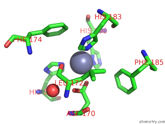
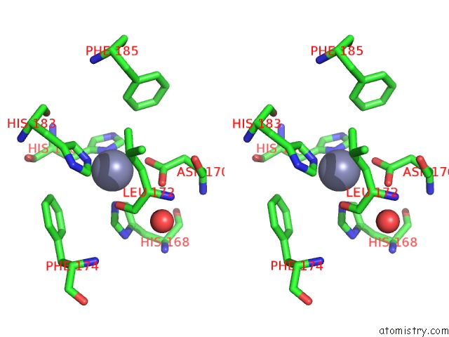
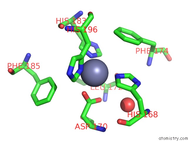
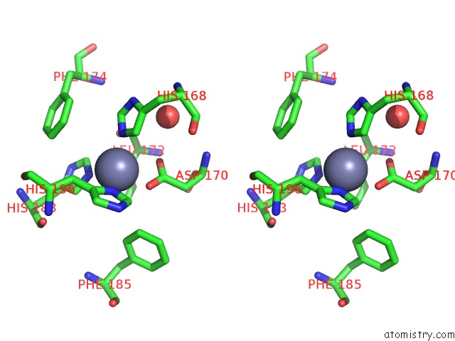
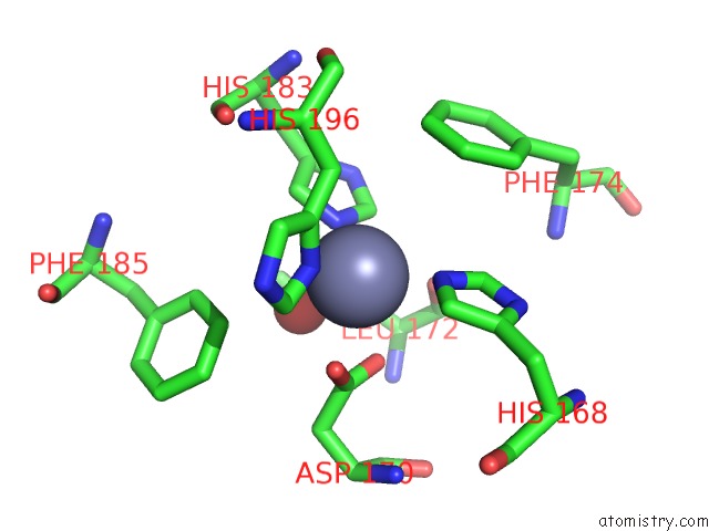
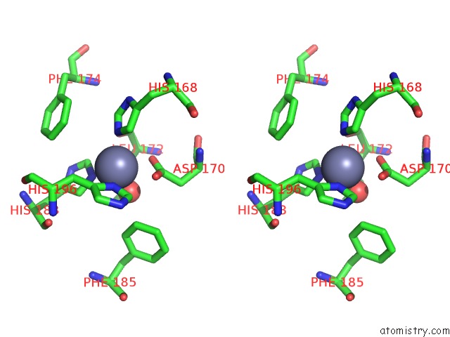
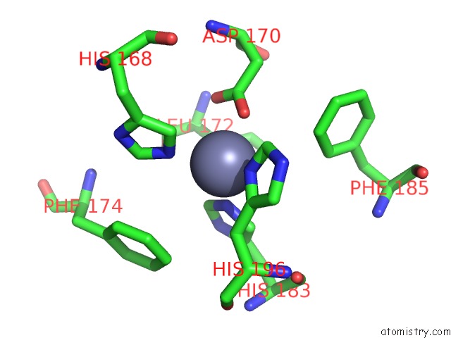
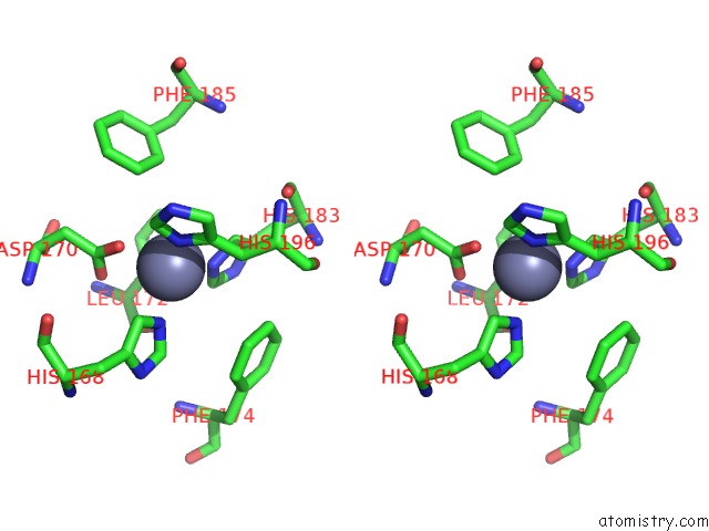
Zinc binding site 1 out of 12 in 1hv5
Go back to
Zinc binding site 1 out
of 12 in the Crystal Structure of the Stromelysin-3 (Mmp-11) Catalytic Domain Complexed with A Phosphinic Inhibitor
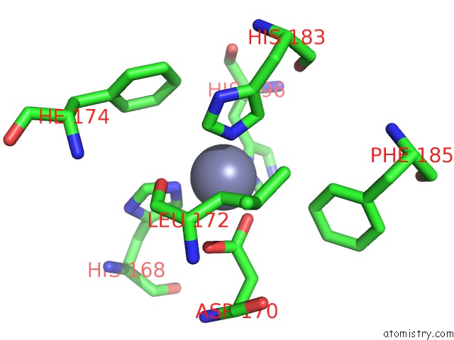
Mono view
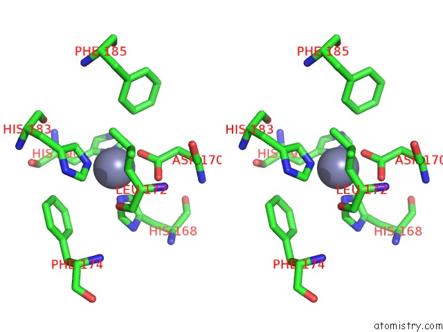
Stereo pair view
Mono view
Stereo pair view
A full contact list of Zinc with other atoms in the Zn binding
site number 1 of Crystal Structure of the Stromelysin-3 (Mmp-11) Catalytic Domain Complexed with A Phosphinic Inhibitor within 5.0Å range:
|
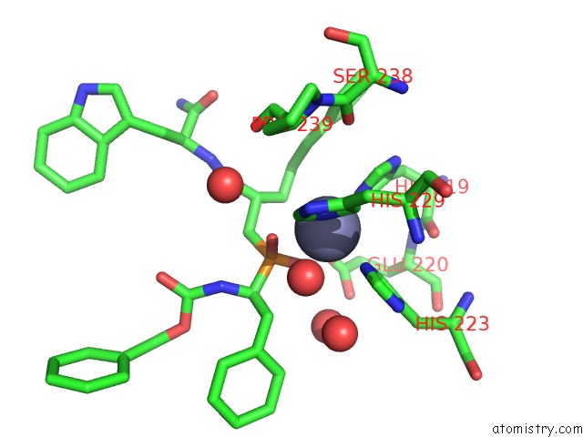
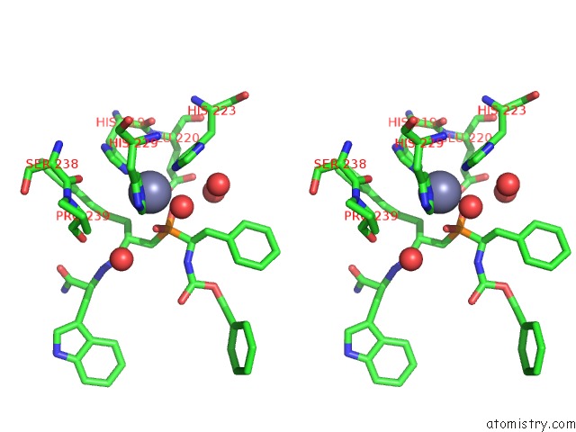
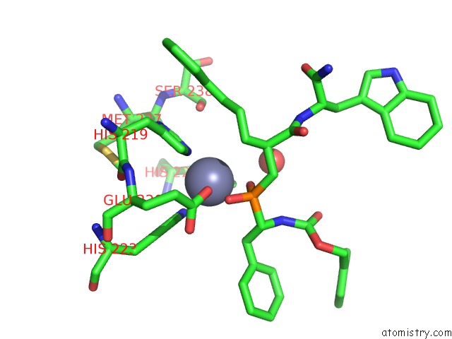
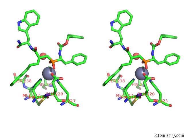
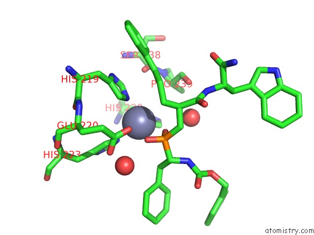
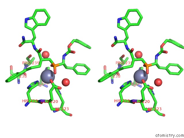
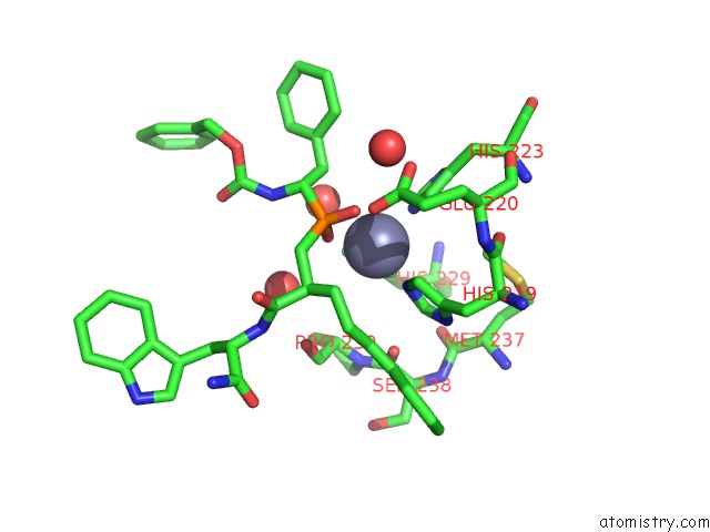
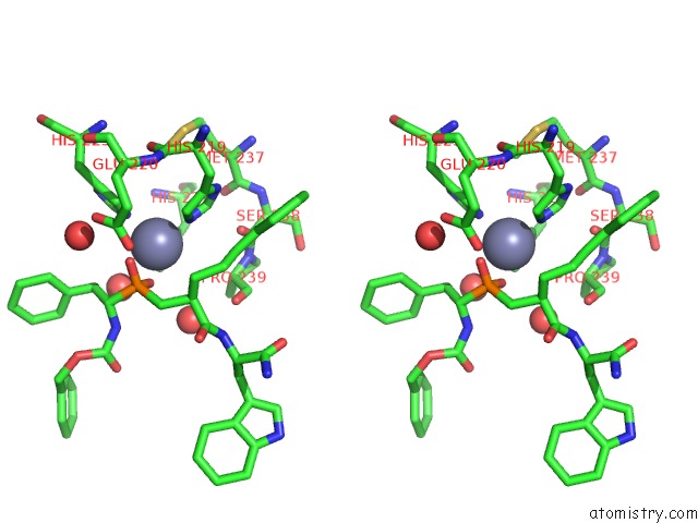
Zinc binding site 2 out of 12 in 1hv5
Go back to
Zinc binding site 2 out
of 12 in the Crystal Structure of the Stromelysin-3 (Mmp-11) Catalytic Domain Complexed with A Phosphinic Inhibitor
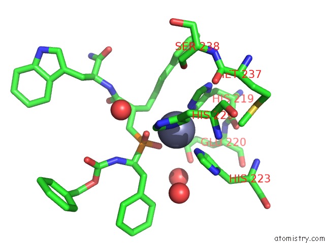
Mono view
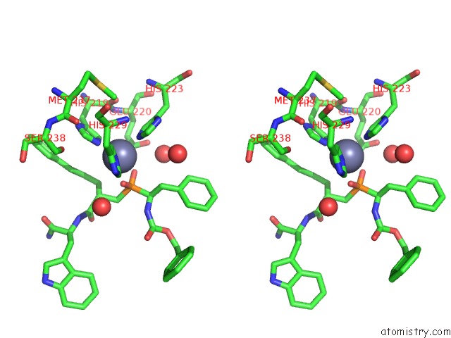
Stereo pair view
Mono view
Stereo pair view
A full contact list of Zinc with other atoms in the Zn binding
site number 2 of Crystal Structure of the Stromelysin-3 (Mmp-11) Catalytic Domain Complexed with A Phosphinic Inhibitor within 5.0Å range:
|
Zinc binding site 3 out of 12 in 1hv5
Go back to
Zinc binding site 3 out
of 12 in the Crystal Structure of the Stromelysin-3 (Mmp-11) Catalytic Domain Complexed with A Phosphinic Inhibitor
Mono view
Stereo pair view
Mono view
Stereo pair view
A full contact list of Zinc with other atoms in the Zn binding
site number 3 of Crystal Structure of the Stromelysin-3 (Mmp-11) Catalytic Domain Complexed with A Phosphinic Inhibitor within 5.0Å range:
|
Zinc binding site 4 out of 12 in 1hv5
Go back to
Zinc binding site 4 out
of 12 in the Crystal Structure of the Stromelysin-3 (Mmp-11) Catalytic Domain Complexed with A Phosphinic Inhibitor
Mono view
Stereo pair view
Mono view
Stereo pair view
A full contact list of Zinc with other atoms in the Zn binding
site number 4 of Crystal Structure of the Stromelysin-3 (Mmp-11) Catalytic Domain Complexed with A Phosphinic Inhibitor within 5.0Å range:
|
Zinc binding site 5 out of 12 in 1hv5
Go back to
Zinc binding site 5 out
of 12 in the Crystal Structure of the Stromelysin-3 (Mmp-11) Catalytic Domain Complexed with A Phosphinic Inhibitor
Mono view
Stereo pair view
Mono view
Stereo pair view
A full contact list of Zinc with other atoms in the Zn binding
site number 5 of Crystal Structure of the Stromelysin-3 (Mmp-11) Catalytic Domain Complexed with A Phosphinic Inhibitor within 5.0Å range:
|
Zinc binding site 6 out of 12 in 1hv5
Go back to
Zinc binding site 6 out
of 12 in the Crystal Structure of the Stromelysin-3 (Mmp-11) Catalytic Domain Complexed with A Phosphinic Inhibitor
Mono view
Stereo pair view
Mono view
Stereo pair view
A full contact list of Zinc with other atoms in the Zn binding
site number 6 of Crystal Structure of the Stromelysin-3 (Mmp-11) Catalytic Domain Complexed with A Phosphinic Inhibitor within 5.0Å range:
|
Zinc binding site 7 out of 12 in 1hv5
Go back to
Zinc binding site 7 out
of 12 in the Crystal Structure of the Stromelysin-3 (Mmp-11) Catalytic Domain Complexed with A Phosphinic Inhibitor
Mono view
Stereo pair view
Mono view
Stereo pair view
A full contact list of Zinc with other atoms in the Zn binding
site number 7 of Crystal Structure of the Stromelysin-3 (Mmp-11) Catalytic Domain Complexed with A Phosphinic Inhibitor within 5.0Å range:
|
Zinc binding site 8 out of 12 in 1hv5
Go back to
Zinc binding site 8 out
of 12 in the Crystal Structure of the Stromelysin-3 (Mmp-11) Catalytic Domain Complexed with A Phosphinic Inhibitor
Mono view
Stereo pair view
Mono view
Stereo pair view
A full contact list of Zinc with other atoms in the Zn binding
site number 8 of Crystal Structure of the Stromelysin-3 (Mmp-11) Catalytic Domain Complexed with A Phosphinic Inhibitor within 5.0Å range:
|
Zinc binding site 9 out of 12 in 1hv5
Go back to
Zinc binding site 9 out
of 12 in the Crystal Structure of the Stromelysin-3 (Mmp-11) Catalytic Domain Complexed with A Phosphinic Inhibitor
Mono view
Stereo pair view
Mono view
Stereo pair view
A full contact list of Zinc with other atoms in the Zn binding
site number 9 of Crystal Structure of the Stromelysin-3 (Mmp-11) Catalytic Domain Complexed with A Phosphinic Inhibitor within 5.0Å range:
|
Zinc binding site 10 out of 12 in 1hv5
Go back to
Zinc binding site 10 out
of 12 in the Crystal Structure of the Stromelysin-3 (Mmp-11) Catalytic Domain Complexed with A Phosphinic Inhibitor
Mono view
Stereo pair view
Mono view
Stereo pair view
A full contact list of Zinc with other atoms in the Zn binding
site number 10 of Crystal Structure of the Stromelysin-3 (Mmp-11) Catalytic Domain Complexed with A Phosphinic Inhibitor within 5.0Å range:
|
Reference:
A.L.Gall,
M.Ruff,
R.Kannan,
P.Cuniasse,
A.Yiotakis,
V.Dive,
M.C.Rio,
P.Basset,
D.Moras.
Crystal Structure of the Stromelysin-3 (Mmp-11) Catalytic Domain Complexed with A Phosphinic Inhibitor Mimicking the Transition-State. J.Mol.Biol. V. 307 577 2001.
ISSN: ISSN 0022-2836
PubMed: 11254383
DOI: 10.1006/JMBI.2001.4493
Page generated: Sun Oct 13 02:35:07 2024
ISSN: ISSN 0022-2836
PubMed: 11254383
DOI: 10.1006/JMBI.2001.4493
Last articles
Zn in 9J0NZn in 9J0O
Zn in 9J0P
Zn in 9FJX
Zn in 9EKB
Zn in 9C0F
Zn in 9CAH
Zn in 9CH0
Zn in 9CH3
Zn in 9CH1