Zinc »
PDB 1gkr-1h4t »
1gr0 »
Zinc in PDB 1gr0: Myo-Inositol 1-Phosphate Synthase From Mycobacterium Tuberculosis in Complex with Nad and Zinc.
Enzymatic activity of Myo-Inositol 1-Phosphate Synthase From Mycobacterium Tuberculosis in Complex with Nad and Zinc.
All present enzymatic activity of Myo-Inositol 1-Phosphate Synthase From Mycobacterium Tuberculosis in Complex with Nad and Zinc.:
5.5.1.4;
5.5.1.4;
Protein crystallography data
The structure of Myo-Inositol 1-Phosphate Synthase From Mycobacterium Tuberculosis in Complex with Nad and Zinc., PDB code: 1gr0
was solved by
R.A.Norman,
J.Murray-Rust,
N.Q.Mcdonald,
Tb Structural Genomicsconsortium (Tbsgc),
with X-Ray Crystallography technique. A brief refinement statistics is given in the table below:
| Resolution Low / High (Å) | 24.79 / 1.95 |
| Space group | P 4 21 2 |
| Cell size a, b, c (Å), α, β, γ (°) | 116.195, 116.195, 64.544, 90.00, 90.00, 90.00 |
| R / Rfree (%) | 21.1 / 23.9 |
Other elements in 1gr0:
The structure of Myo-Inositol 1-Phosphate Synthase From Mycobacterium Tuberculosis in Complex with Nad and Zinc. also contains other interesting chemical elements:
| Arsenic | (As) | 1 atom |
Zinc Binding Sites:
The binding sites of Zinc atom in the Myo-Inositol 1-Phosphate Synthase From Mycobacterium Tuberculosis in Complex with Nad and Zinc.
(pdb code 1gr0). This binding sites where shown within
5.0 Angstroms radius around Zinc atom.
In total only one binding site of Zinc was determined in the Myo-Inositol 1-Phosphate Synthase From Mycobacterium Tuberculosis in Complex with Nad and Zinc., PDB code: 1gr0:
In total only one binding site of Zinc was determined in the Myo-Inositol 1-Phosphate Synthase From Mycobacterium Tuberculosis in Complex with Nad and Zinc., PDB code: 1gr0:
Zinc binding site 1 out of 1 in 1gr0
Go back to
Zinc binding site 1 out
of 1 in the Myo-Inositol 1-Phosphate Synthase From Mycobacterium Tuberculosis in Complex with Nad and Zinc.
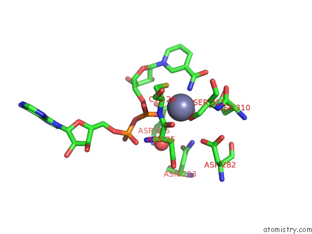
Mono view
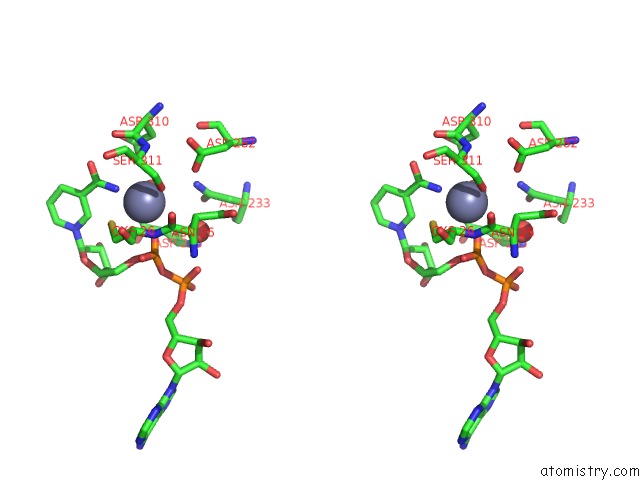
Stereo pair view
Mono view
Stereo pair view
A full contact list of Zinc with other atoms in the Zn binding
site number 1 of Myo-Inositol 1-Phosphate Synthase From Mycobacterium Tuberculosis in Complex with Nad and Zinc. within 5.0Å range:
|
Reference:
R.A.Norman,
M.S.B.Mcalister,
J.Murray-Rust,
F.Movahedzadeh,
N.G.Stoker,
N.Q.Mcdonald.
Crystal Structure of Inositol 1-Phosphate Synthase From Mycobacterium Tuberculosis, A Key Enzyme in Phosphatidylinositol Synthesis Structure V. 10 393 2002.
ISSN: ISSN 0969-2126
PubMed: 12005437
DOI: 10.1016/S0969-2126(02)00718-9
Page generated: Sun Oct 13 01:40:40 2024
ISSN: ISSN 0969-2126
PubMed: 12005437
DOI: 10.1016/S0969-2126(02)00718-9
Last articles
Zn in 9J0NZn in 9J0O
Zn in 9J0P
Zn in 9FJX
Zn in 9EKB
Zn in 9C0F
Zn in 9CAH
Zn in 9CH0
Zn in 9CH3
Zn in 9CH1