Zinc »
PDB 1fpp-1g43 »
1ft1 »
Zinc in PDB 1ft1: Crystal Structure of Protein Farnesyltransferase at 2.25 Angstroms Resolution
Protein crystallography data
The structure of Crystal Structure of Protein Farnesyltransferase at 2.25 Angstroms Resolution, PDB code: 1ft1
was solved by
L.S.Beese,
H.-W.Park,
with X-Ray Crystallography technique. A brief refinement statistics is given in the table below:
| Resolution Low / High (Å) | 6.00 / 2.25 |
| Space group | P 65 |
| Cell size a, b, c (Å), α, β, γ (°) | 167.060, 167.060, 97.900, 90.00, 90.00, 120.00 |
| R / Rfree (%) | 21 / 26 |
Zinc Binding Sites:
The binding sites of Zinc atom in the Crystal Structure of Protein Farnesyltransferase at 2.25 Angstroms Resolution
(pdb code 1ft1). This binding sites where shown within
5.0 Angstroms radius around Zinc atom.
In total only one binding site of Zinc was determined in the Crystal Structure of Protein Farnesyltransferase at 2.25 Angstroms Resolution, PDB code: 1ft1:
In total only one binding site of Zinc was determined in the Crystal Structure of Protein Farnesyltransferase at 2.25 Angstroms Resolution, PDB code: 1ft1:
Zinc binding site 1 out of 1 in 1ft1
Go back to
Zinc binding site 1 out
of 1 in the Crystal Structure of Protein Farnesyltransferase at 2.25 Angstroms Resolution
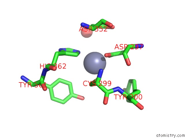
Mono view
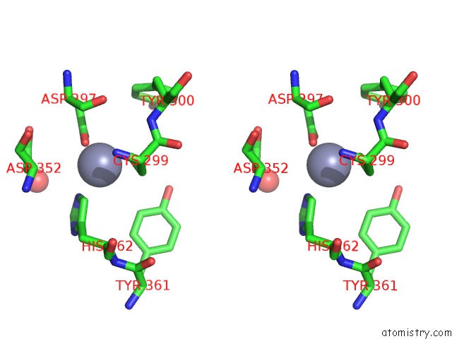
Stereo pair view
Mono view
Stereo pair view
A full contact list of Zinc with other atoms in the Zn binding
site number 1 of Crystal Structure of Protein Farnesyltransferase at 2.25 Angstroms Resolution within 5.0Å range:
|
Reference:
H.W.Park,
S.R.Boduluri,
J.F.Moomaw,
P.J.Casey,
L.S.Beese.
Crystal Structure of Protein Farnesyltransferase at 2.25 Angstrom Resolution. Science V. 275 1800 1997.
ISSN: ISSN 0036-8075
PubMed: 9065406
DOI: 10.1126/SCIENCE.275.5307.1800
Page generated: Sun Oct 13 01:07:59 2024
ISSN: ISSN 0036-8075
PubMed: 9065406
DOI: 10.1126/SCIENCE.275.5307.1800
Last articles
Zn in 9J0NZn in 9J0O
Zn in 9J0P
Zn in 9FJX
Zn in 9EKB
Zn in 9C0F
Zn in 9CAH
Zn in 9CH0
Zn in 9CH3
Zn in 9CH1