Zinc »
PDB 1ed8-1evl »
1ev6 »
Zinc in PDB 1ev6: Structure of the Monoclinic Form of the M-Cresol/Insulin R6 Hexamer
Protein crystallography data
The structure of Structure of the Monoclinic Form of the M-Cresol/Insulin R6 Hexamer, PDB code: 1ev6
was solved by
G.D.Smith,
E.Ciszak,
L.A.Magrum,
W.A.Pangborn,
R.H.Blessing,
with X-Ray Crystallography technique. A brief refinement statistics is given in the table below:
| Resolution Low / High (Å) | 20.95 / 1.90 |
| Space group | P 1 21 1 |
| Cell size a, b, c (Å), α, β, γ (°) | 61.247, 61.739, 47.467, 90.00, 111.32, 90.00 |
| R / Rfree (%) | 19.5 / 23.5 |
Other elements in 1ev6:
The structure of Structure of the Monoclinic Form of the M-Cresol/Insulin R6 Hexamer also contains other interesting chemical elements:
| Chlorine | (Cl) | 2 atoms |
| Sodium | (Na) | 1 atom |
Zinc Binding Sites:
The binding sites of Zinc atom in the Structure of the Monoclinic Form of the M-Cresol/Insulin R6 Hexamer
(pdb code 1ev6). This binding sites where shown within
5.0 Angstroms radius around Zinc atom.
In total 2 binding sites of Zinc where determined in the Structure of the Monoclinic Form of the M-Cresol/Insulin R6 Hexamer, PDB code: 1ev6:
Jump to Zinc binding site number: 1; 2;
In total 2 binding sites of Zinc where determined in the Structure of the Monoclinic Form of the M-Cresol/Insulin R6 Hexamer, PDB code: 1ev6:
Jump to Zinc binding site number: 1; 2;
Zinc binding site 1 out of 2 in 1ev6
Go back to
Zinc binding site 1 out
of 2 in the Structure of the Monoclinic Form of the M-Cresol/Insulin R6 Hexamer
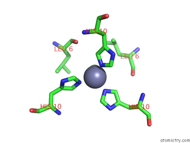
Mono view
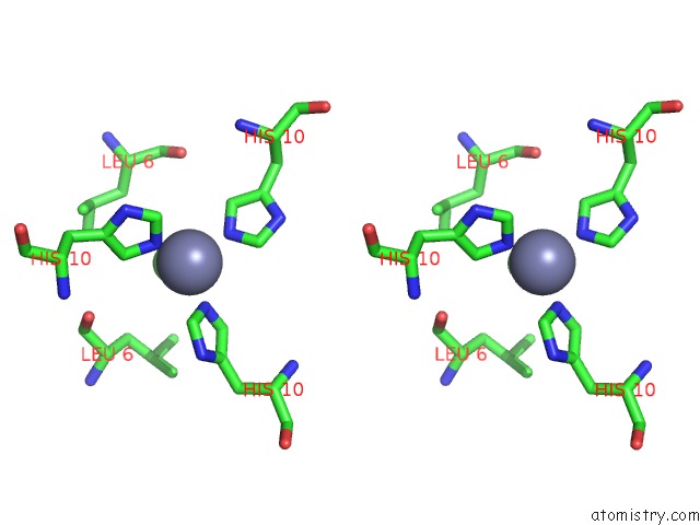
Stereo pair view
Mono view
Stereo pair view
A full contact list of Zinc with other atoms in the Zn binding
site number 1 of Structure of the Monoclinic Form of the M-Cresol/Insulin R6 Hexamer within 5.0Å range:
|
Zinc binding site 2 out of 2 in 1ev6
Go back to
Zinc binding site 2 out
of 2 in the Structure of the Monoclinic Form of the M-Cresol/Insulin R6 Hexamer
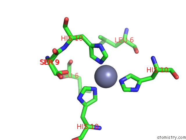
Mono view
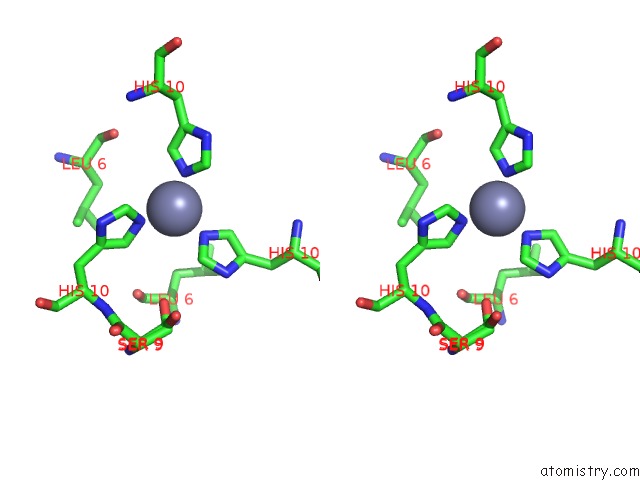
Stereo pair view
Mono view
Stereo pair view
A full contact list of Zinc with other atoms in the Zn binding
site number 2 of Structure of the Monoclinic Form of the M-Cresol/Insulin R6 Hexamer within 5.0Å range:
|
Reference:
G.D.Smith,
E.Ciszak,
L.A.Magrum,
W.A.Pangborn,
R.H.Blessing.
R6 Hexameric Insulin Complexed with M-Cresol or Resorcinol Biochem.Biophys.Res.Commun. V. 56 1541 2000.
ISSN: ISSN 0006-291X
PubMed: 11092919
DOI: 10.1107/S0907444900012749
Page generated: Sun Oct 13 00:25:51 2024
ISSN: ISSN 0006-291X
PubMed: 11092919
DOI: 10.1107/S0907444900012749
Last articles
Zn in 9J0NZn in 9J0O
Zn in 9J0P
Zn in 9FJX
Zn in 9EKB
Zn in 9C0F
Zn in 9CAH
Zn in 9CH0
Zn in 9CH3
Zn in 9CH1