Zinc »
PDB 1ed8-1evl »
1eu4 »
Zinc in PDB 1eu4: Crystal Structure of the Superantigen Spe-H (Zinc Bound) From Streptococcus Pyogenes
Protein crystallography data
The structure of Crystal Structure of the Superantigen Spe-H (Zinc Bound) From Streptococcus Pyogenes, PDB code: 1eu4
was solved by
V.L.Arcus,
T.Proft,
J.A.Sigrell,
H.M.Baker,
J.D.Fraser,
E.N.Baker,
with X-Ray Crystallography technique. A brief refinement statistics is given in the table below:
| Resolution Low / High (Å) | 19.49 / 2.50 |
| Space group | P 1 21 1 |
| Cell size a, b, c (Å), α, β, γ (°) | 36.629, 46.028, 64.470, 90.00, 91.64, 90.00 |
| R / Rfree (%) | 20.5 / 25.8 |
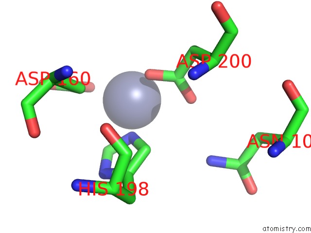
Zinc Binding Sites:
The binding sites of Zinc atom in the Crystal Structure of the Superantigen Spe-H (Zinc Bound) From Streptococcus Pyogenes
(pdb code 1eu4). This binding sites where shown within
5.0 Angstroms radius around Zinc atom.
In total 2 binding sites of Zinc where determined in the Crystal Structure of the Superantigen Spe-H (Zinc Bound) From Streptococcus Pyogenes, PDB code: 1eu4:
Jump to Zinc binding site number: 1; 2;
In total 2 binding sites of Zinc where determined in the Crystal Structure of the Superantigen Spe-H (Zinc Bound) From Streptococcus Pyogenes, PDB code: 1eu4:
Jump to Zinc binding site number: 1; 2;
Zinc binding site 1 out of 2 in 1eu4
Go back to
Zinc binding site 1 out
of 2 in the Crystal Structure of the Superantigen Spe-H (Zinc Bound) From Streptococcus Pyogenes
Mono view

Stereo pair view
Mono view
Stereo pair view
A full contact list of Zinc with other atoms in the Zn binding
site number 1 of Crystal Structure of the Superantigen Spe-H (Zinc Bound) From Streptococcus Pyogenes within 5.0Å range:
|
Zinc binding site 2 out of 2 in 1eu4
Go back to
Zinc binding site 2 out
of 2 in the Crystal Structure of the Superantigen Spe-H (Zinc Bound) From Streptococcus Pyogenes

Mono view

Stereo pair view
Mono view
Stereo pair view
A full contact list of Zinc with other atoms in the Zn binding
site number 2 of Crystal Structure of the Superantigen Spe-H (Zinc Bound) From Streptococcus Pyogenes within 5.0Å range:
|
Reference:
V.L.Arcus,
T.Proft,
J.A.Sigrell,
H.M.Baker,
J.D.Fraser,
E.N.Baker.
Conservation and Variation in Superantigen Structure and Activity Highlighted By the Three-Dimensional Structures of Two New Superantigens From Streptococcus Pyogenes. J.Mol.Biol. V. 299 157 2000.
ISSN: ISSN 0022-2836
PubMed: 10860729
DOI: 10.1006/JMBI.2000.3725
Page generated: Sun Oct 13 00:24:43 2024
ISSN: ISSN 0022-2836
PubMed: 10860729
DOI: 10.1006/JMBI.2000.3725
Last articles
Zn in 9J0NZn in 9J0O
Zn in 9J0P
Zn in 9FJX
Zn in 9EKB
Zn in 9C0F
Zn in 9CAH
Zn in 9CH0
Zn in 9CH3
Zn in 9CH1