Zinc »
PDB 1caq-1co4 »
1cni »
Zinc in PDB 1cni: X-Ray Crystallographic Studies of Engineered Hydrogen Bond Networks in A Protein-Zinc Binding Site
Enzymatic activity of X-Ray Crystallographic Studies of Engineered Hydrogen Bond Networks in A Protein-Zinc Binding Site
All present enzymatic activity of X-Ray Crystallographic Studies of Engineered Hydrogen Bond Networks in A Protein-Zinc Binding Site:
4.2.1.1;
4.2.1.1;
Protein crystallography data
The structure of X-Ray Crystallographic Studies of Engineered Hydrogen Bond Networks in A Protein-Zinc Binding Site, PDB code: 1cni
was solved by
C.A.Lesburg,
D.W.Christianson,
with X-Ray Crystallography technique. A brief refinement statistics is given in the table below:
| Resolution Low / High (Å) | 8.00 / 1.80 |
| Space group | P 1 21 1 |
| Cell size a, b, c (Å), α, β, γ (°) | 42.700, 41.700, 73.000, 90.00, 104.60, 90.00 |
| R / Rfree (%) | 16.1 / n/a |
Other elements in 1cni:
The structure of X-Ray Crystallographic Studies of Engineered Hydrogen Bond Networks in A Protein-Zinc Binding Site also contains other interesting chemical elements:
| Mercury | (Hg) | 1 atom |
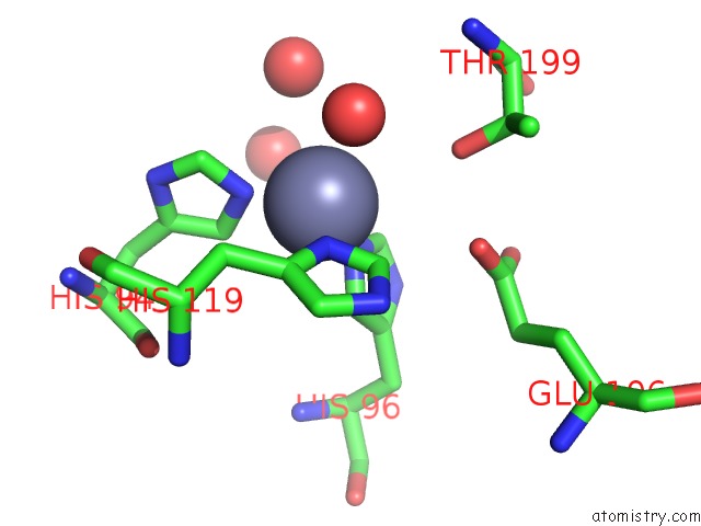
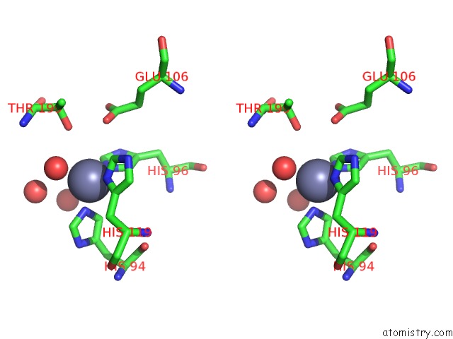
Zinc Binding Sites:
The binding sites of Zinc atom in the X-Ray Crystallographic Studies of Engineered Hydrogen Bond Networks in A Protein-Zinc Binding Site
(pdb code 1cni). This binding sites where shown within
5.0 Angstroms radius around Zinc atom.
In total only one binding site of Zinc was determined in the X-Ray Crystallographic Studies of Engineered Hydrogen Bond Networks in A Protein-Zinc Binding Site, PDB code: 1cni:
In total only one binding site of Zinc was determined in the X-Ray Crystallographic Studies of Engineered Hydrogen Bond Networks in A Protein-Zinc Binding Site, PDB code: 1cni:
Zinc binding site 1 out of 1 in 1cni
Go back to
Zinc binding site 1 out
of 1 in the X-Ray Crystallographic Studies of Engineered Hydrogen Bond Networks in A Protein-Zinc Binding Site
Mono view
Stereo pair view
Mono view
Stereo pair view
A full contact list of Zinc with other atoms in the Zn binding
site number 1 of X-Ray Crystallographic Studies of Engineered Hydrogen Bond Networks in A Protein-Zinc Binding Site within 5.0Å range:
|
Reference:
C.A.Lesburg,
D.W.Christianson.
X-Ray Crystallographic Studies of Engineered Hydrogen Bond Networks in A Protein-Zinc Binding Site J.Am.Chem.Soc. V. 117 6838 1995.
ISSN: ISSN 0002-7863
Page generated: Sat Oct 12 23:12:44 2024
ISSN: ISSN 0002-7863
Last articles
Zn in 9J0NZn in 9J0O
Zn in 9J0P
Zn in 9FJX
Zn in 9EKB
Zn in 9C0F
Zn in 9CAH
Zn in 9CH0
Zn in 9CH3
Zn in 9CH1