Zinc »
PDB 1adf-1axg »
1ajb »
Zinc in PDB 1ajb: Three-Dimensional Structure of the D153G Mutant of E. Coli Alkaline Phosphatase: A Mutant with Weaker Magnesium Binding and Increased Catalytic Activity
Enzymatic activity of Three-Dimensional Structure of the D153G Mutant of E. Coli Alkaline Phosphatase: A Mutant with Weaker Magnesium Binding and Increased Catalytic Activity
All present enzymatic activity of Three-Dimensional Structure of the D153G Mutant of E. Coli Alkaline Phosphatase: A Mutant with Weaker Magnesium Binding and Increased Catalytic Activity:
3.1.3.1;
3.1.3.1;
Protein crystallography data
The structure of Three-Dimensional Structure of the D153G Mutant of E. Coli Alkaline Phosphatase: A Mutant with Weaker Magnesium Binding and Increased Catalytic Activity, PDB code: 1ajb
was solved by
C.G.Dealwis,
L.Chen,
C.Abad-Zapatero,
with X-Ray Crystallography technique. A brief refinement statistics is given in the table below:
| Resolution Low / High (Å) | 6.00 / 2.50 |
| Space group | I 2 2 2 |
| Cell size a, b, c (Å), α, β, γ (°) | 195.020, 166.930, 76.440, 90.00, 90.00, 90.00 |
| R / Rfree (%) | 16.2 / n/a |
Other elements in 1ajb:
The structure of Three-Dimensional Structure of the D153G Mutant of E. Coli Alkaline Phosphatase: A Mutant with Weaker Magnesium Binding and Increased Catalytic Activity also contains other interesting chemical elements:
| Magnesium | (Mg) | 2 atoms |
Zinc Binding Sites:
The binding sites of Zinc atom in the Three-Dimensional Structure of the D153G Mutant of E. Coli Alkaline Phosphatase: A Mutant with Weaker Magnesium Binding and Increased Catalytic Activity
(pdb code 1ajb). This binding sites where shown within
5.0 Angstroms radius around Zinc atom.
In total 4 binding sites of Zinc where determined in the Three-Dimensional Structure of the D153G Mutant of E. Coli Alkaline Phosphatase: A Mutant with Weaker Magnesium Binding and Increased Catalytic Activity, PDB code: 1ajb:
Jump to Zinc binding site number: 1; 2; 3; 4;
In total 4 binding sites of Zinc where determined in the Three-Dimensional Structure of the D153G Mutant of E. Coli Alkaline Phosphatase: A Mutant with Weaker Magnesium Binding and Increased Catalytic Activity, PDB code: 1ajb:
Jump to Zinc binding site number: 1; 2; 3; 4;
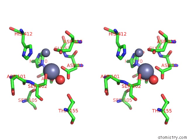
Zinc binding site 1 out of 4 in 1ajb
Go back to
Zinc binding site 1 out
of 4 in the Three-Dimensional Structure of the D153G Mutant of E. Coli Alkaline Phosphatase: A Mutant with Weaker Magnesium Binding and Increased Catalytic Activity

Mono view
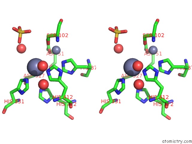
Stereo pair view
Mono view
Stereo pair view
A full contact list of Zinc with other atoms in the Zn binding
site number 1 of Three-Dimensional Structure of the D153G Mutant of E. Coli Alkaline Phosphatase: A Mutant with Weaker Magnesium Binding and Increased Catalytic Activity within 5.0Å range:
|
Zinc binding site 2 out of 4 in 1ajb
Go back to
Zinc binding site 2 out
of 4 in the Three-Dimensional Structure of the D153G Mutant of E. Coli Alkaline Phosphatase: A Mutant with Weaker Magnesium Binding and Increased Catalytic Activity

Mono view
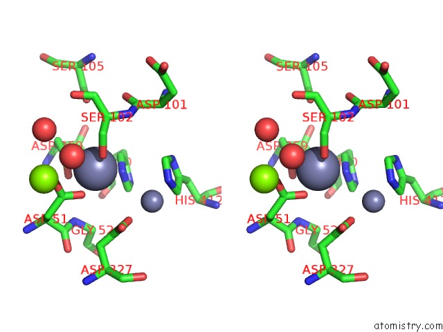
Stereo pair view
Mono view
Stereo pair view
A full contact list of Zinc with other atoms in the Zn binding
site number 2 of Three-Dimensional Structure of the D153G Mutant of E. Coli Alkaline Phosphatase: A Mutant with Weaker Magnesium Binding and Increased Catalytic Activity within 5.0Å range:
|
Zinc binding site 3 out of 4 in 1ajb
Go back to
Zinc binding site 3 out
of 4 in the Three-Dimensional Structure of the D153G Mutant of E. Coli Alkaline Phosphatase: A Mutant with Weaker Magnesium Binding and Increased Catalytic Activity
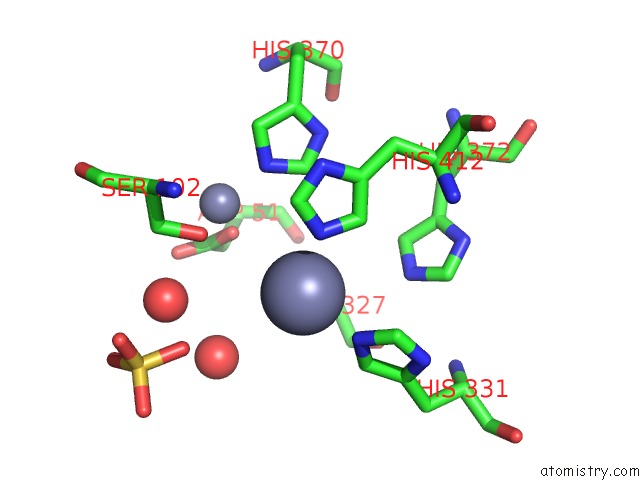
Mono view

Stereo pair view
Mono view
Stereo pair view
A full contact list of Zinc with other atoms in the Zn binding
site number 3 of Three-Dimensional Structure of the D153G Mutant of E. Coli Alkaline Phosphatase: A Mutant with Weaker Magnesium Binding and Increased Catalytic Activity within 5.0Å range:
|
Zinc binding site 4 out of 4 in 1ajb
Go back to
Zinc binding site 4 out
of 4 in the Three-Dimensional Structure of the D153G Mutant of E. Coli Alkaline Phosphatase: A Mutant with Weaker Magnesium Binding and Increased Catalytic Activity
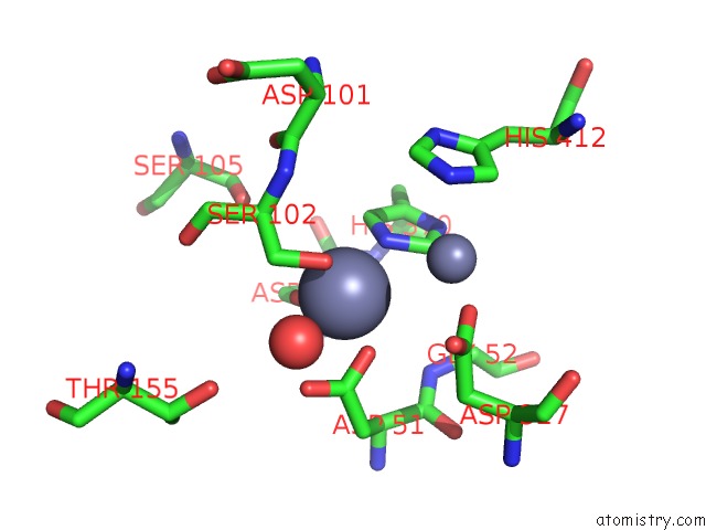
Mono view
Stereo pair view
Mono view
Stereo pair view
A full contact list of Zinc with other atoms in the Zn binding
site number 4 of Three-Dimensional Structure of the D153G Mutant of E. Coli Alkaline Phosphatase: A Mutant with Weaker Magnesium Binding and Increased Catalytic Activity within 5.0Å range:
|
Reference:
C.G.Dealwis,
L.Chen,
C.Brennan,
W.Mandecki,
C.Abad-Zapatero.
3-D Structure of the D153G Mutant of Escherichia Coli Alkaline Phosphatase: An Enzyme with Weaker Magnesium Binding and Increased Catalytic Activity. Protein Eng. V. 8 865 1995.
ISSN: ISSN 0269-2139
PubMed: 8746724
DOI: 10.1093/PROTEIN/8.9.865
Page generated: Sat Oct 12 22:03:18 2024
ISSN: ISSN 0269-2139
PubMed: 8746724
DOI: 10.1093/PROTEIN/8.9.865
Last articles
Zn in 9J0NZn in 9J0O
Zn in 9J0P
Zn in 9FJX
Zn in 9EKB
Zn in 9C0F
Zn in 9CAH
Zn in 9CH0
Zn in 9CH3
Zn in 9CH1