Zinc »
PDB 12ca-1add »
1a6f »
Zinc in PDB 1a6f: Rnase P Protein From Bacillus Subtilis
Enzymatic activity of Rnase P Protein From Bacillus Subtilis
All present enzymatic activity of Rnase P Protein From Bacillus Subtilis:
3.1.26.5;
3.1.26.5;
Protein crystallography data
The structure of Rnase P Protein From Bacillus Subtilis, PDB code: 1a6f
was solved by
T.Stams,
D.W.Christianson,
with X-Ray Crystallography technique. A brief refinement statistics is given in the table below:
| Resolution Low / High (Å) | 20.00 / 2.60 |
| Space group | P 64 |
| Cell size a, b, c (Å), α, β, γ (°) | 82.400, 82.400, 33.500, 90.00, 90.00, 120.00 |
| R / Rfree (%) | 20.5 / 31.7 |
Zinc Binding Sites:
The binding sites of Zinc atom in the Rnase P Protein From Bacillus Subtilis
(pdb code 1a6f). This binding sites where shown within
5.0 Angstroms radius around Zinc atom.
In total 2 binding sites of Zinc where determined in the Rnase P Protein From Bacillus Subtilis, PDB code: 1a6f:
Jump to Zinc binding site number: 1; 2;
In total 2 binding sites of Zinc where determined in the Rnase P Protein From Bacillus Subtilis, PDB code: 1a6f:
Jump to Zinc binding site number: 1; 2;
Zinc binding site 1 out of 2 in 1a6f
Go back to
Zinc binding site 1 out
of 2 in the Rnase P Protein From Bacillus Subtilis
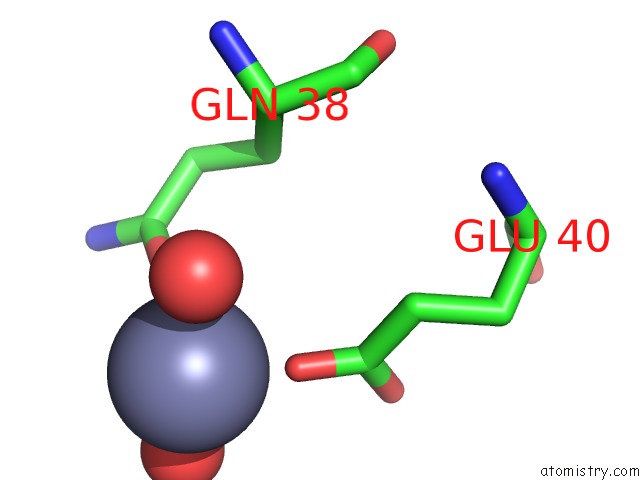
Mono view
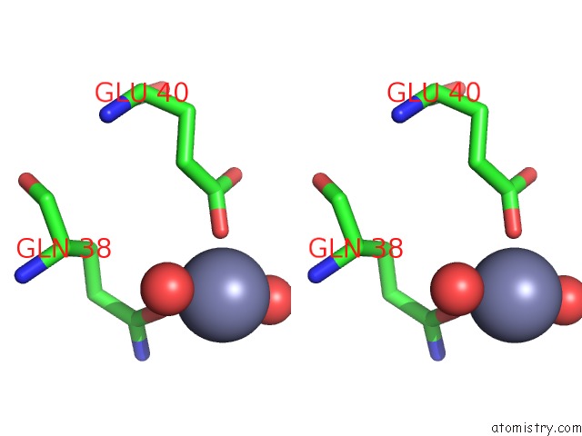
Stereo pair view
Mono view
Stereo pair view
A full contact list of Zinc with other atoms in the Zn binding
site number 1 of Rnase P Protein From Bacillus Subtilis within 5.0Å range:
|
Zinc binding site 2 out of 2 in 1a6f
Go back to
Zinc binding site 2 out
of 2 in the Rnase P Protein From Bacillus Subtilis
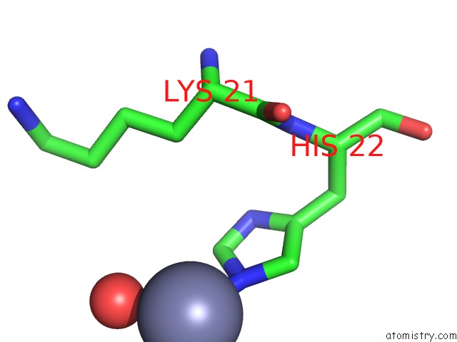
Mono view
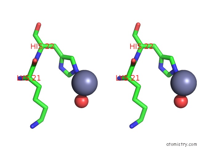
Stereo pair view
Mono view
Stereo pair view
A full contact list of Zinc with other atoms in the Zn binding
site number 2 of Rnase P Protein From Bacillus Subtilis within 5.0Å range:
|
Reference:
T.Stams,
S.Niranjanakumari,
C.A.Fierke,
D.W.Christianson.
Ribonuclease P Protein Structure: Evolutionary Origins in the Translational Apparatus. Science V. 280 752 1998.
ISSN: ISSN 0036-8075
PubMed: 9563955
DOI: 10.1126/SCIENCE.280.5364.752
Page generated: Sat Oct 12 21:48:09 2024
ISSN: ISSN 0036-8075
PubMed: 9563955
DOI: 10.1126/SCIENCE.280.5364.752
Last articles
Zn in 9J0NZn in 9J0O
Zn in 9J0P
Zn in 9FJX
Zn in 9EKB
Zn in 9C0F
Zn in 9CAH
Zn in 9CH0
Zn in 9CH3
Zn in 9CH1