Zinc »
PDB 9f3p-9fne »
9fk6 »
Zinc in PDB 9fk6: The Structure of XT6 From G.Proteiniphilus T-6: the E265G/N158D Mutant
Enzymatic activity of The Structure of XT6 From G.Proteiniphilus T-6: the E265G/N158D Mutant
All present enzymatic activity of The Structure of XT6 From G.Proteiniphilus T-6: the E265G/N158D Mutant:
3.2.1.8;
3.2.1.8;
Protein crystallography data
The structure of The Structure of XT6 From G.Proteiniphilus T-6: the E265G/N158D Mutant, PDB code: 9fk6
was solved by
N.Hadad,
O.Chmelnik,
M.Dessau,
Y.Shoham,
G.Shoham,
with X-Ray Crystallography technique. A brief refinement statistics is given in the table below:
| Resolution Low / High (Å) | 36.55 / 1.95 |
| Space group | I 1 2 1 |
| Cell size a, b, c (Å), α, β, γ (°) | 89.133, 61.874, 110.956, 90, 105.37, 90 |
| R / Rfree (%) | 17.7 / 21.1 |
Other elements in 9fk6:
The structure of The Structure of XT6 From G.Proteiniphilus T-6: the E265G/N158D Mutant also contains other interesting chemical elements:
| Chlorine | (Cl) | 2 atoms |
Zinc Binding Sites:
The binding sites of Zinc atom in the The Structure of XT6 From G.Proteiniphilus T-6: the E265G/N158D Mutant
(pdb code 9fk6). This binding sites where shown within
5.0 Angstroms radius around Zinc atom.
In total 7 binding sites of Zinc where determined in the The Structure of XT6 From G.Proteiniphilus T-6: the E265G/N158D Mutant, PDB code: 9fk6:
Jump to Zinc binding site number: 1; 2; 3; 4; 5; 6; 7;
In total 7 binding sites of Zinc where determined in the The Structure of XT6 From G.Proteiniphilus T-6: the E265G/N158D Mutant, PDB code: 9fk6:
Jump to Zinc binding site number: 1; 2; 3; 4; 5; 6; 7;
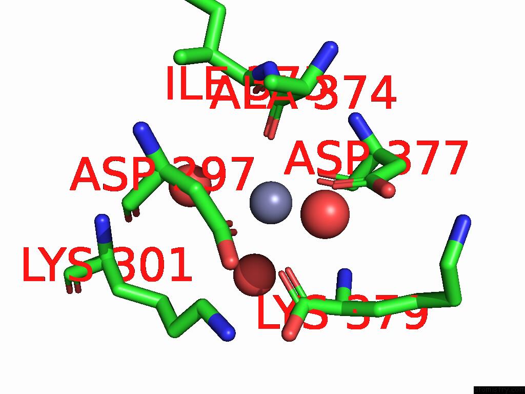
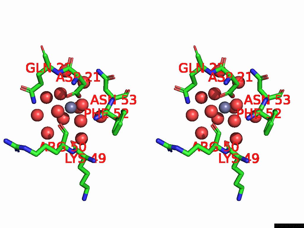
Zinc binding site 1 out of 7 in 9fk6
Go back to
Zinc binding site 1 out
of 7 in the The Structure of XT6 From G.Proteiniphilus T-6: the E265G/N158D Mutant
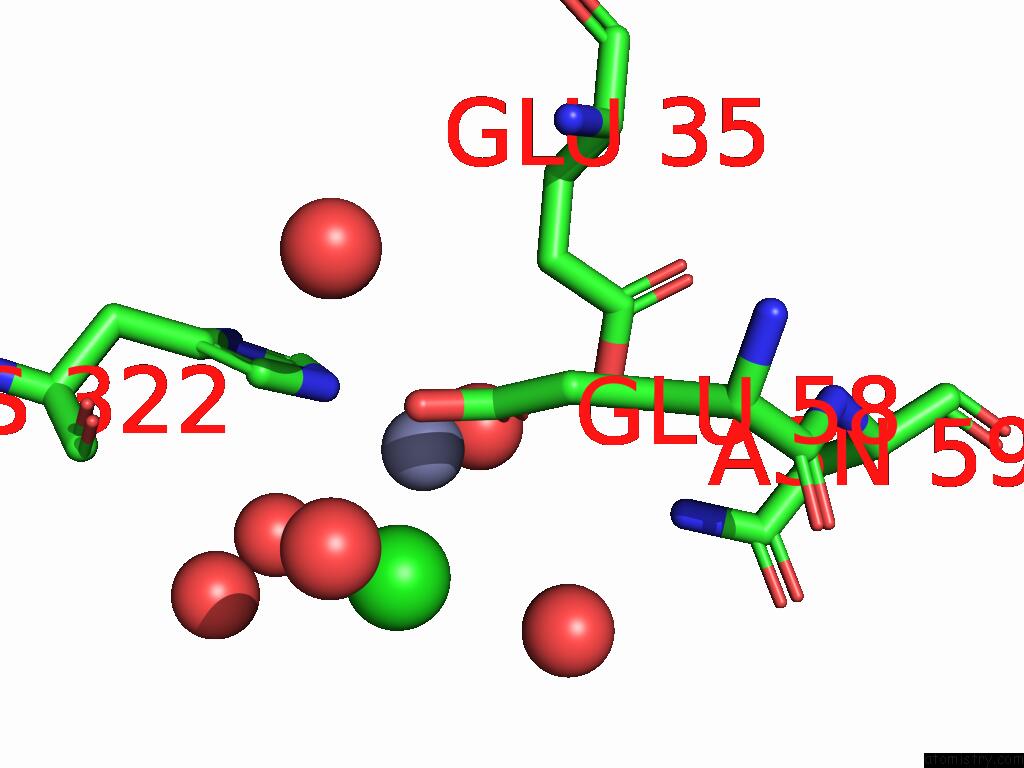
Mono view
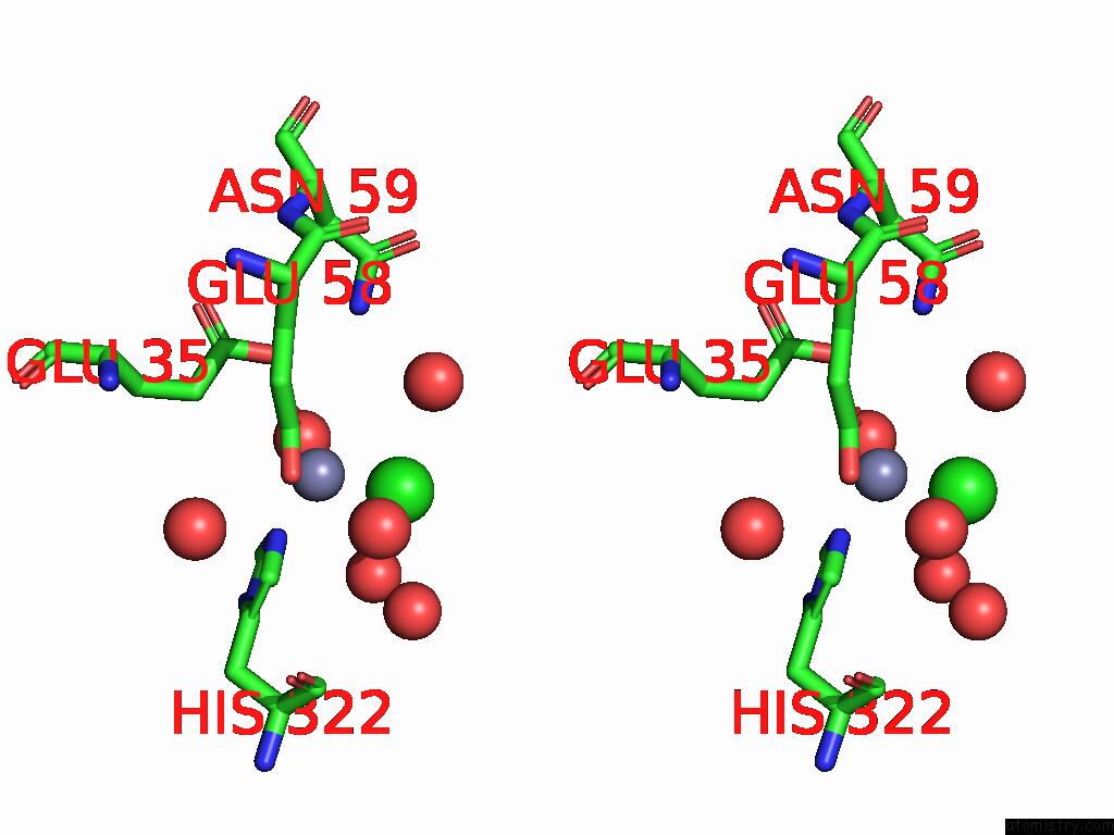
Stereo pair view
Mono view
Stereo pair view
A full contact list of Zinc with other atoms in the Zn binding
site number 1 of The Structure of XT6 From G.Proteiniphilus T-6: the E265G/N158D Mutant within 5.0Å range:
|
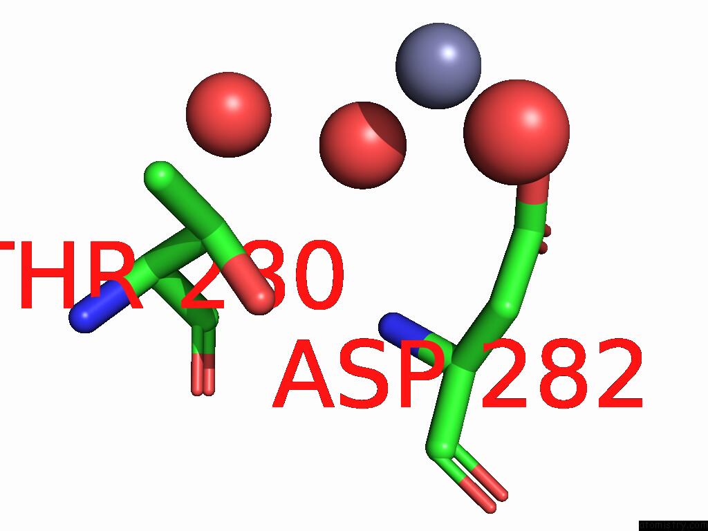
Zinc binding site 2 out of 7 in 9fk6
Go back to
Zinc binding site 2 out
of 7 in the The Structure of XT6 From G.Proteiniphilus T-6: the E265G/N158D Mutant
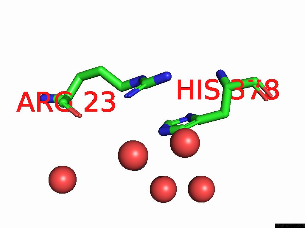
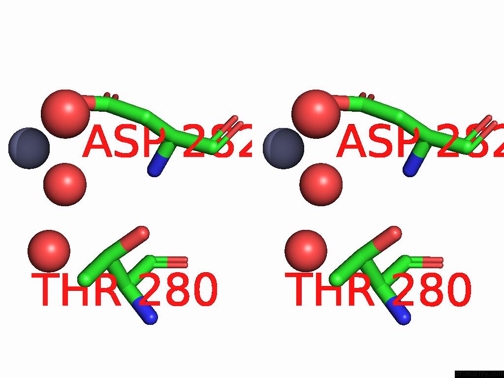
Mono view
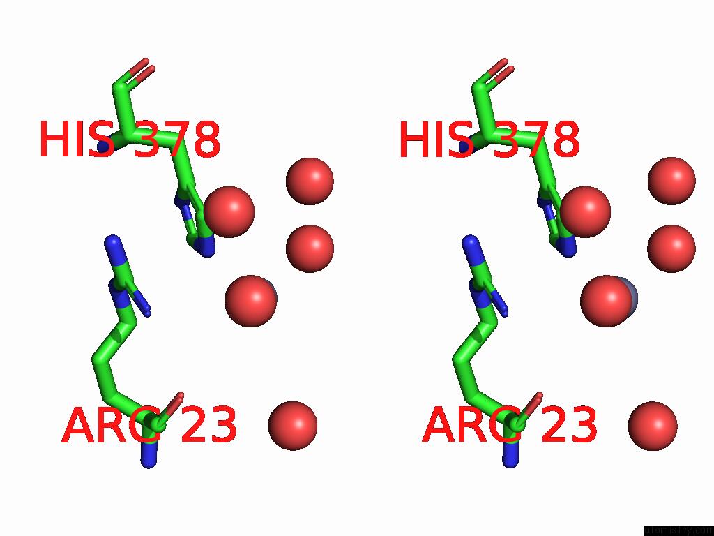
Stereo pair view
Mono view
Stereo pair view
A full contact list of Zinc with other atoms in the Zn binding
site number 2 of The Structure of XT6 From G.Proteiniphilus T-6: the E265G/N158D Mutant within 5.0Å range:
|
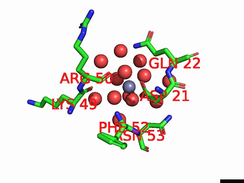
Zinc binding site 3 out of 7 in 9fk6
Go back to
Zinc binding site 3 out
of 7 in the The Structure of XT6 From G.Proteiniphilus T-6: the E265G/N158D Mutant
Mono view

Stereo pair view
Mono view
Stereo pair view
A full contact list of Zinc with other atoms in the Zn binding
site number 3 of The Structure of XT6 From G.Proteiniphilus T-6: the E265G/N158D Mutant within 5.0Å range:
|
Zinc binding site 4 out of 7 in 9fk6
Go back to
Zinc binding site 4 out
of 7 in the The Structure of XT6 From G.Proteiniphilus T-6: the E265G/N158D Mutant
Mono view
Stereo pair view
Mono view
Stereo pair view
A full contact list of Zinc with other atoms in the Zn binding
site number 4 of The Structure of XT6 From G.Proteiniphilus T-6: the E265G/N158D Mutant within 5.0Å range:
|
Zinc binding site 5 out of 7 in 9fk6
Go back to
Zinc binding site 5 out
of 7 in the The Structure of XT6 From G.Proteiniphilus T-6: the E265G/N158D Mutant

Mono view

Stereo pair view
Mono view
Stereo pair view
A full contact list of Zinc with other atoms in the Zn binding
site number 5 of The Structure of XT6 From G.Proteiniphilus T-6: the E265G/N158D Mutant within 5.0Å range:
|
Zinc binding site 6 out of 7 in 9fk6
Go back to
Zinc binding site 6 out
of 7 in the The Structure of XT6 From G.Proteiniphilus T-6: the E265G/N158D Mutant
Mono view
Stereo pair view
Mono view
Stereo pair view
A full contact list of Zinc with other atoms in the Zn binding
site number 6 of The Structure of XT6 From G.Proteiniphilus T-6: the E265G/N158D Mutant within 5.0Å range:
|
Zinc binding site 7 out of 7 in 9fk6
Go back to
Zinc binding site 7 out
of 7 in the The Structure of XT6 From G.Proteiniphilus T-6: the E265G/N158D Mutant

Mono view

Stereo pair view
Mono view
Stereo pair view
A full contact list of Zinc with other atoms in the Zn binding
site number 7 of The Structure of XT6 From G.Proteiniphilus T-6: the E265G/N158D Mutant within 5.0Å range:
|
Reference:
N.Hadad,
O.Chmelnik,
M.Dessau,
Y.Shoham,
G.Shoham.
The Structure of XT6 From G.Proteiniphilus T-6: the E265G/N158D Mutant To Be Published.
Page generated: Fri Aug 22 17:31:12 2025
Last articles
Br in 9R0QBr in 9J73
Br in 9BJ5
Br in 8Y72
Au in 9D33
As in 9O9I
Al in 9GSG
Zr in 1XC1
Zr in 6Y7P
Zr in 6GNL