Zinc »
PDB 9dch-9ed0 »
9dih »
Zinc in PDB 9dih: Cbass Pseudomonas Syringae CAP5 Tetramer with Dna Duplex and 3'2'-C- Diamp Cyclic Dinucleotide Ligand
Protein crystallography data
The structure of Cbass Pseudomonas Syringae CAP5 Tetramer with Dna Duplex and 3'2'-C- Diamp Cyclic Dinucleotide Ligand, PDB code: 9dih
was solved by
O.Rechkoblit,
A.K.Aggarwal,
with X-Ray Crystallography technique. A brief refinement statistics is given in the table below:
| Resolution Low / High (Å) | 47.88 / 1.94 |
| Space group | C 2 2 21 |
| Cell size a, b, c (Å), α, β, γ (°) | 103.697, 191.527, 118.435, 90, 90, 90 |
| R / Rfree (%) | 17.1 / 21.8 |
Zinc Binding Sites:
The binding sites of Zinc atom in the Cbass Pseudomonas Syringae CAP5 Tetramer with Dna Duplex and 3'2'-C- Diamp Cyclic Dinucleotide Ligand
(pdb code 9dih). This binding sites where shown within
5.0 Angstroms radius around Zinc atom.
In total 2 binding sites of Zinc where determined in the Cbass Pseudomonas Syringae CAP5 Tetramer with Dna Duplex and 3'2'-C- Diamp Cyclic Dinucleotide Ligand, PDB code: 9dih:
Jump to Zinc binding site number: 1; 2;
In total 2 binding sites of Zinc where determined in the Cbass Pseudomonas Syringae CAP5 Tetramer with Dna Duplex and 3'2'-C- Diamp Cyclic Dinucleotide Ligand, PDB code: 9dih:
Jump to Zinc binding site number: 1; 2;
Zinc binding site 1 out of 2 in 9dih
Go back to
Zinc binding site 1 out
of 2 in the Cbass Pseudomonas Syringae CAP5 Tetramer with Dna Duplex and 3'2'-C- Diamp Cyclic Dinucleotide Ligand
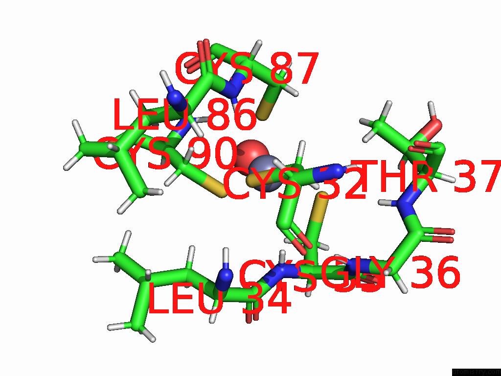
Mono view
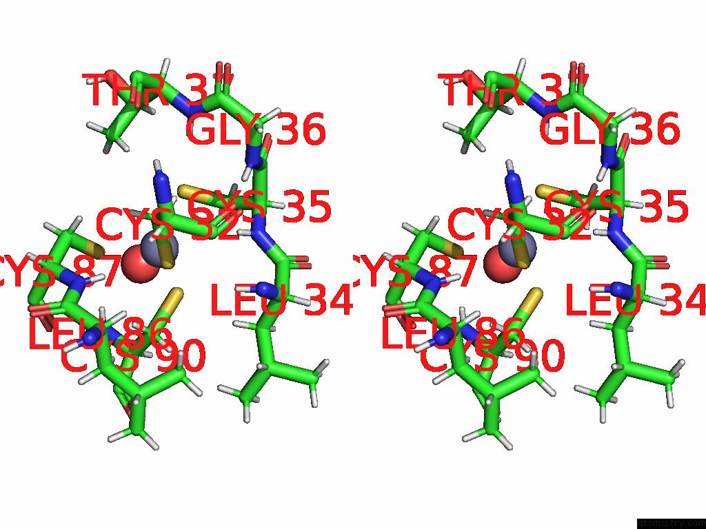
Stereo pair view
Mono view
Stereo pair view
A full contact list of Zinc with other atoms in the Zn binding
site number 1 of Cbass Pseudomonas Syringae CAP5 Tetramer with Dna Duplex and 3'2'-C- Diamp Cyclic Dinucleotide Ligand within 5.0Å range:
|
Zinc binding site 2 out of 2 in 9dih
Go back to
Zinc binding site 2 out
of 2 in the Cbass Pseudomonas Syringae CAP5 Tetramer with Dna Duplex and 3'2'-C- Diamp Cyclic Dinucleotide Ligand
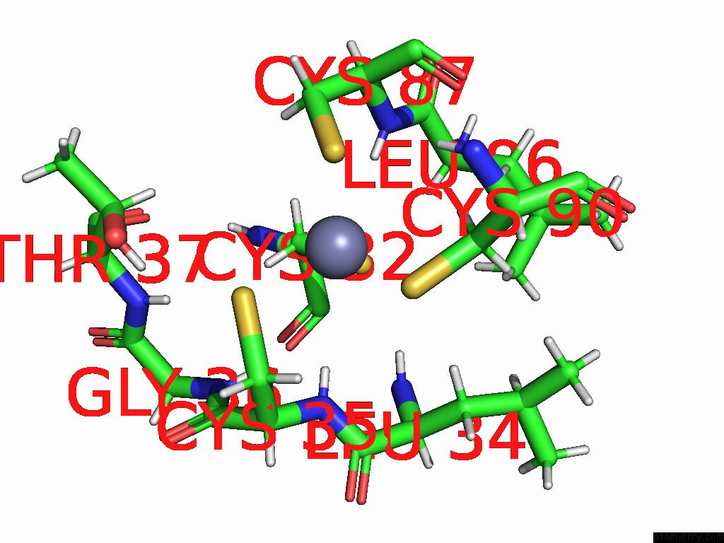
Mono view
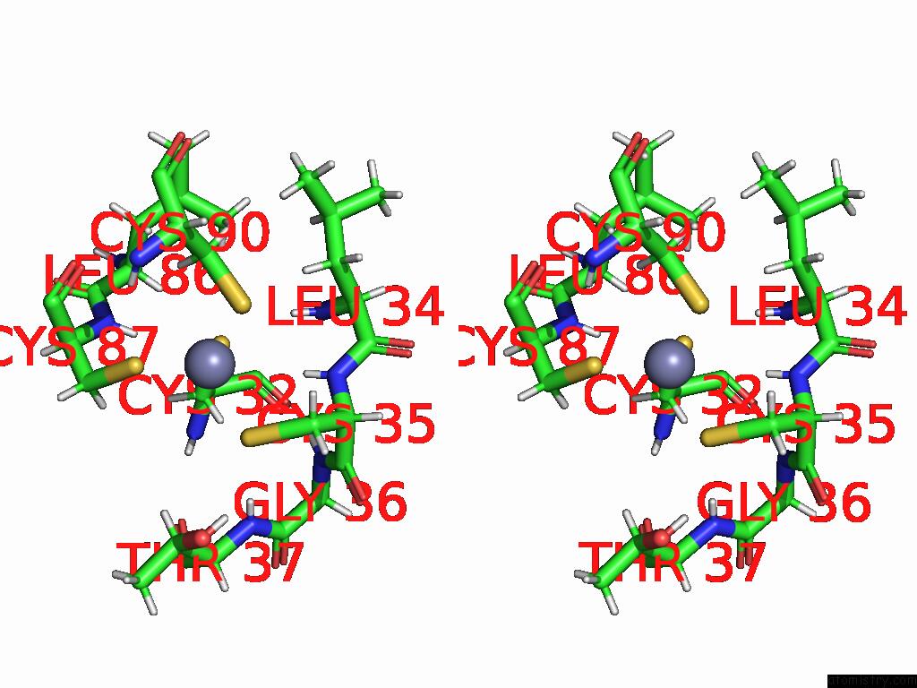
Stereo pair view
Mono view
Stereo pair view
A full contact list of Zinc with other atoms in the Zn binding
site number 2 of Cbass Pseudomonas Syringae CAP5 Tetramer with Dna Duplex and 3'2'-C- Diamp Cyclic Dinucleotide Ligand within 5.0Å range:
|
Reference:
O.Rechkoblit,
D.Sciaky,
M.Ni,
Y.Li,
J.Kottur,
G.Fang,
A.K.Aggarwal.
Mechanism of Dna Degradation By Cbass CAP5 Endonuclease Immune Effector. Nat Commun V. 16 5243 2025.
ISSN: ESSN 2041-1723
PubMed: 40473611
DOI: 10.1038/S41467-025-60484-Z
Page generated: Fri Aug 22 17:07:39 2025
ISSN: ESSN 2041-1723
PubMed: 40473611
DOI: 10.1038/S41467-025-60484-Z
Last articles
Br in 9R0QBr in 9J73
Br in 9BJ5
Br in 8Y72
Au in 9D33
As in 9O9I
Al in 9GSG
Zr in 1XC1
Zr in 6Y7P
Zr in 6GNL