Zinc »
PDB 8zj5-9bap »
9ayu »
Zinc in PDB 9ayu: Structure of the A Type Blood Alpha-D-Galactosamine Galactosaminidase D463A Mutant From Flavonifractor Plautii
Protein crystallography data
The structure of Structure of the A Type Blood Alpha-D-Galactosamine Galactosaminidase D463A Mutant From Flavonifractor Plautii, PDB code: 9ayu
was solved by
L.J.Worrall,
N.C.J.Strynadka,
with X-Ray Crystallography technique. A brief refinement statistics is given in the table below:
| Resolution Low / High (Å) | 48.90 / 2.00 |
| Space group | P 1 21 1 |
| Cell size a, b, c (Å), α, β, γ (°) | 102.326, 164.29, 131.763, 90, 90.2, 90 |
| R / Rfree (%) | 16.3 / 18.6 |
Other elements in 9ayu:
The structure of Structure of the A Type Blood Alpha-D-Galactosamine Galactosaminidase D463A Mutant From Flavonifractor Plautii also contains other interesting chemical elements:
| Chlorine | (Cl) | 5 atoms |
| Manganese | (Mn) | 5 atoms |
Zinc Binding Sites:
The binding sites of Zinc atom in the Structure of the A Type Blood Alpha-D-Galactosamine Galactosaminidase D463A Mutant From Flavonifractor Plautii
(pdb code 9ayu). This binding sites where shown within
5.0 Angstroms radius around Zinc atom.
In total 5 binding sites of Zinc where determined in the Structure of the A Type Blood Alpha-D-Galactosamine Galactosaminidase D463A Mutant From Flavonifractor Plautii, PDB code: 9ayu:
Jump to Zinc binding site number: 1; 2; 3; 4; 5;
In total 5 binding sites of Zinc where determined in the Structure of the A Type Blood Alpha-D-Galactosamine Galactosaminidase D463A Mutant From Flavonifractor Plautii, PDB code: 9ayu:
Jump to Zinc binding site number: 1; 2; 3; 4; 5;
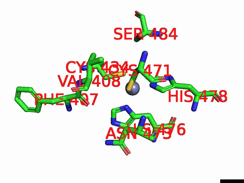
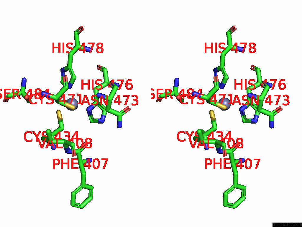
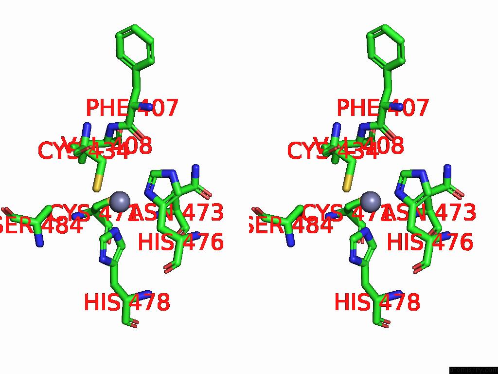
Zinc binding site 1 out of 5 in 9ayu
Go back to
Zinc binding site 1 out
of 5 in the Structure of the A Type Blood Alpha-D-Galactosamine Galactosaminidase D463A Mutant From Flavonifractor Plautii
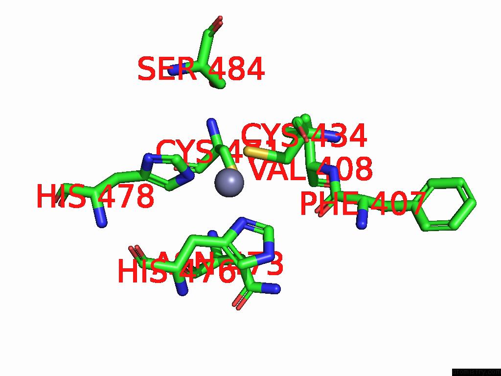
Mono view
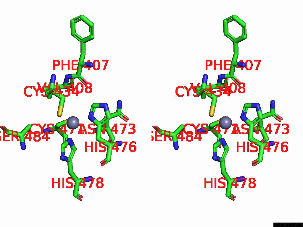
Stereo pair view
Mono view
Stereo pair view
A full contact list of Zinc with other atoms in the Zn binding
site number 1 of Structure of the A Type Blood Alpha-D-Galactosamine Galactosaminidase D463A Mutant From Flavonifractor Plautii within 5.0Å range:
|
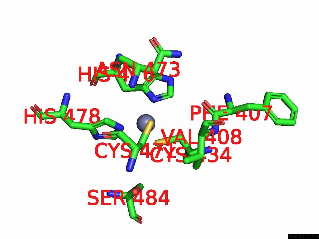
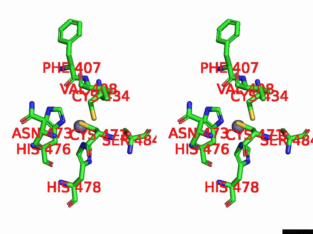
Zinc binding site 2 out of 5 in 9ayu
Go back to
Zinc binding site 2 out
of 5 in the Structure of the A Type Blood Alpha-D-Galactosamine Galactosaminidase D463A Mutant From Flavonifractor Plautii
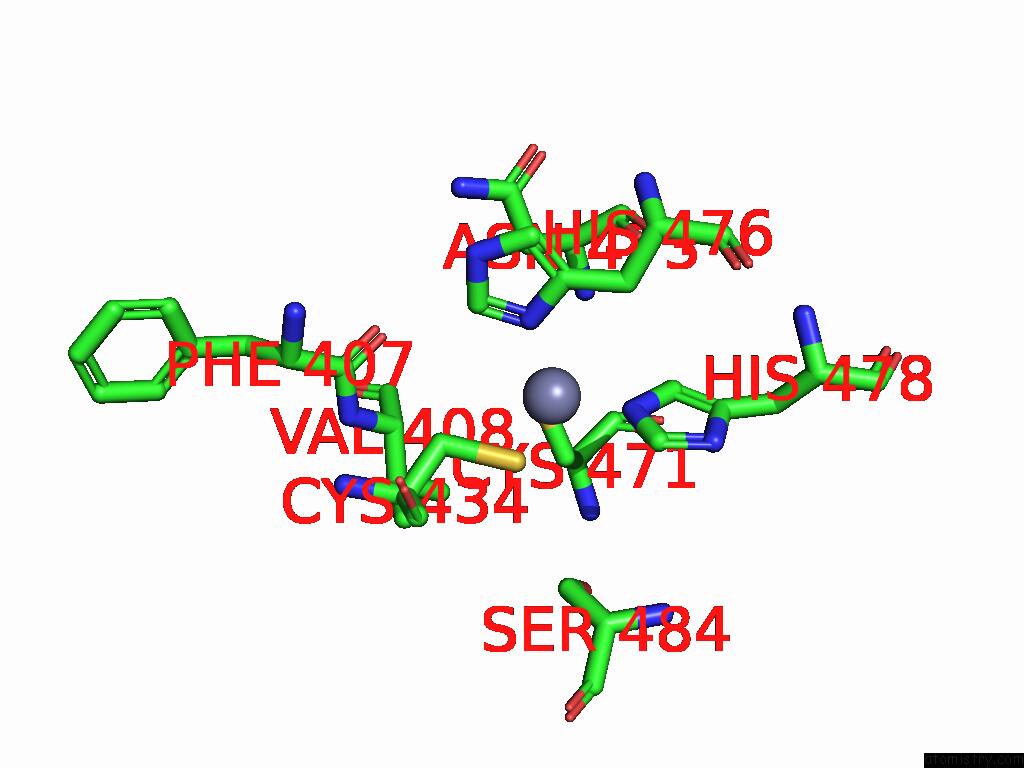
Mono view
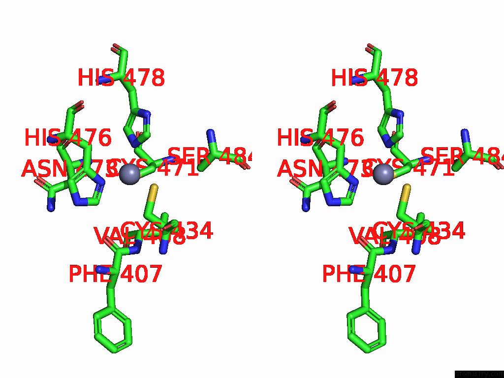
Stereo pair view
Mono view
Stereo pair view
A full contact list of Zinc with other atoms in the Zn binding
site number 2 of Structure of the A Type Blood Alpha-D-Galactosamine Galactosaminidase D463A Mutant From Flavonifractor Plautii within 5.0Å range:
|
Zinc binding site 3 out of 5 in 9ayu
Go back to
Zinc binding site 3 out
of 5 in the Structure of the A Type Blood Alpha-D-Galactosamine Galactosaminidase D463A Mutant From Flavonifractor Plautii
Mono view
Stereo pair view
Mono view
Stereo pair view
A full contact list of Zinc with other atoms in the Zn binding
site number 3 of Structure of the A Type Blood Alpha-D-Galactosamine Galactosaminidase D463A Mutant From Flavonifractor Plautii within 5.0Å range:
|
Zinc binding site 4 out of 5 in 9ayu
Go back to
Zinc binding site 4 out
of 5 in the Structure of the A Type Blood Alpha-D-Galactosamine Galactosaminidase D463A Mutant From Flavonifractor Plautii
Mono view
Stereo pair view
Mono view
Stereo pair view
A full contact list of Zinc with other atoms in the Zn binding
site number 4 of Structure of the A Type Blood Alpha-D-Galactosamine Galactosaminidase D463A Mutant From Flavonifractor Plautii within 5.0Å range:
|
Zinc binding site 5 out of 5 in 9ayu
Go back to
Zinc binding site 5 out
of 5 in the Structure of the A Type Blood Alpha-D-Galactosamine Galactosaminidase D463A Mutant From Flavonifractor Plautii

Mono view
Stereo pair view
Mono view
Stereo pair view
A full contact list of Zinc with other atoms in the Zn binding
site number 5 of Structure of the A Type Blood Alpha-D-Galactosamine Galactosaminidase D463A Mutant From Flavonifractor Plautii within 5.0Å range:
|
Reference:
Y.Tian,
L.J.Worrall,
L.Sim,
F.Liu,
S.A.Nasseri,
P.Rahfeld,
W.Mu,
J.N.Kizhakkedathu,
N.C.J.Strynadka,
S.G.Withers.
Cobalt As A Cofactor For Alpha-Galactosaminidase-Catalyzed Cleavage of Blood Group Antigens Acs Catalysis 2024.
ISSN: ESSN 2155-5435
DOI: 10.1021/ACSCATAL.4C03643
Page generated: Sun Feb 9 01:00:26 2025
ISSN: ESSN 2155-5435
DOI: 10.1021/ACSCATAL.4C03643
Last articles
Na in 6JZINa in 6JYV
Na in 6JZH
Na in 6JY4
Na in 6JY3
Na in 6JUW
Na in 6JPR
Na in 6JSC
Na in 6JSG
Na in 6JNH