Zinc »
PDB 8vne-8w34 »
8vud »
Zinc in PDB 8vud: Crystal Structure of APOBEC3F-CD1
Protein crystallography data
The structure of Crystal Structure of APOBEC3F-CD1, PDB code: 8vud
was solved by
H.J.Yang,
S.-X.Li,
J.Pacheco,
X.S.Chen,
with X-Ray Crystallography technique. A brief refinement statistics is given in the table below:
| Resolution Low / High (Å) | 41.21 / 2.60 |
| Space group | P 6 |
| Cell size a, b, c (Å), α, β, γ (°) | 124.388, 124.388, 64.011, 90, 90, 120 |
| R / Rfree (%) | 17.8 / 23.9 |
Zinc Binding Sites:
The binding sites of Zinc atom in the Crystal Structure of APOBEC3F-CD1
(pdb code 8vud). This binding sites where shown within
5.0 Angstroms radius around Zinc atom.
In total 4 binding sites of Zinc where determined in the Crystal Structure of APOBEC3F-CD1, PDB code: 8vud:
Jump to Zinc binding site number: 1; 2; 3; 4;
In total 4 binding sites of Zinc where determined in the Crystal Structure of APOBEC3F-CD1, PDB code: 8vud:
Jump to Zinc binding site number: 1; 2; 3; 4;
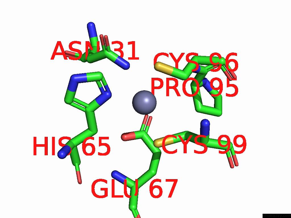
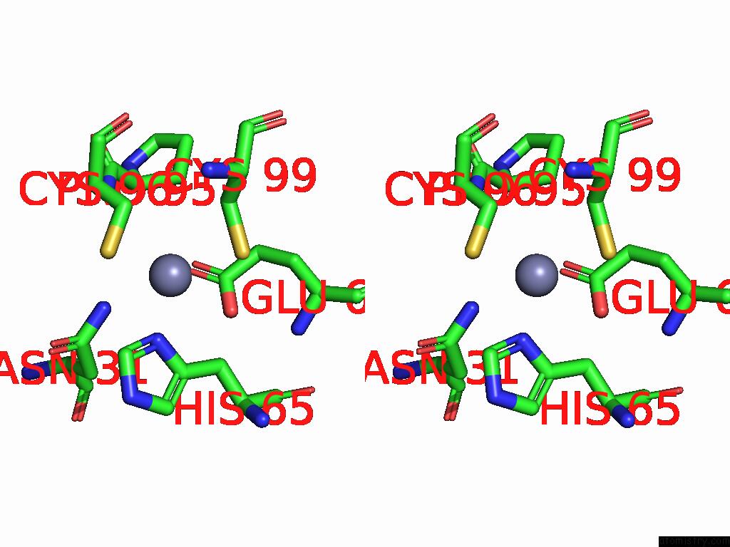
Zinc binding site 1 out of 4 in 8vud
Go back to
Zinc binding site 1 out
of 4 in the Crystal Structure of APOBEC3F-CD1
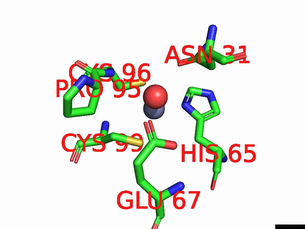
Mono view
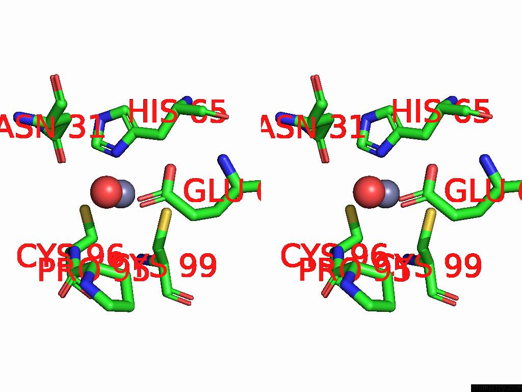
Stereo pair view
Mono view
Stereo pair view
A full contact list of Zinc with other atoms in the Zn binding
site number 1 of Crystal Structure of APOBEC3F-CD1 within 5.0Å range:
|
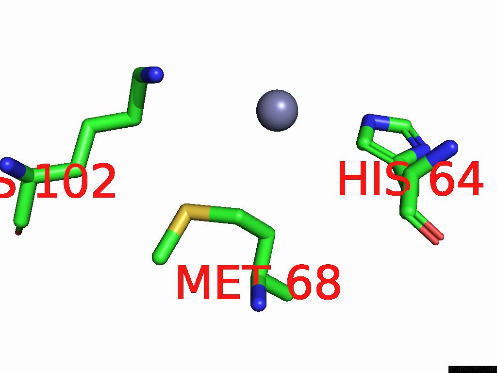
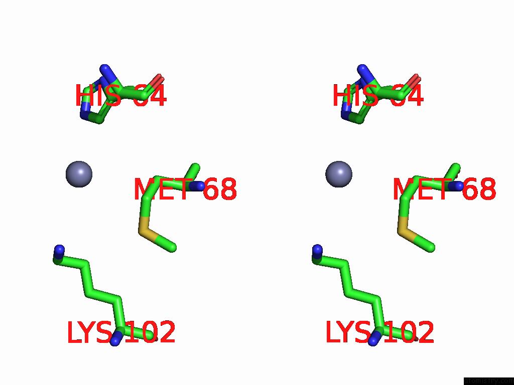
Zinc binding site 2 out of 4 in 8vud
Go back to
Zinc binding site 2 out
of 4 in the Crystal Structure of APOBEC3F-CD1
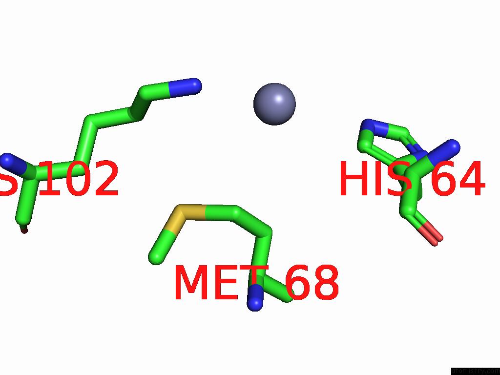
Mono view
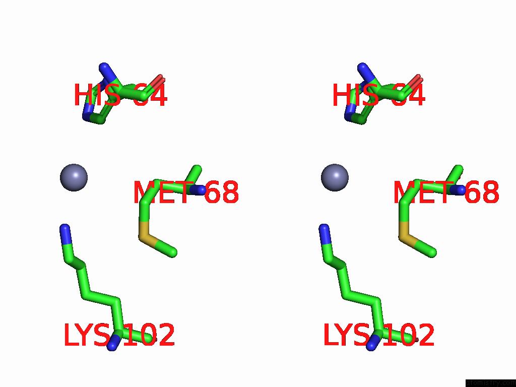
Stereo pair view
Mono view
Stereo pair view
A full contact list of Zinc with other atoms in the Zn binding
site number 2 of Crystal Structure of APOBEC3F-CD1 within 5.0Å range:
|
Zinc binding site 3 out of 4 in 8vud
Go back to
Zinc binding site 3 out
of 4 in the Crystal Structure of APOBEC3F-CD1
Mono view
Stereo pair view
Mono view
Stereo pair view
A full contact list of Zinc with other atoms in the Zn binding
site number 3 of Crystal Structure of APOBEC3F-CD1 within 5.0Å range:
|
Zinc binding site 4 out of 4 in 8vud
Go back to
Zinc binding site 4 out
of 4 in the Crystal Structure of APOBEC3F-CD1
Mono view
Stereo pair view
Mono view
Stereo pair view
A full contact list of Zinc with other atoms in the Zn binding
site number 4 of Crystal Structure of APOBEC3F-CD1 within 5.0Å range:
|
Reference:
J.Pacheco,
H.J.Yang,
S.-X.Li,
X.S.Chen.
Crystal Structure of APOBEC3F-CD1 To Be Published.
Page generated: Fri Aug 22 14:53:28 2025
Last articles
Zr in 1XC1Zr in 6Y7P
Zr in 6GNL
Zr in 6HYB
Zr in 4XYY
Zr in 5KHP
Zn in 9VXG
Zn in 9VWY
Zn in 9VCL
Zn in 9VKN