Zinc »
PDB 6tm9-6ty2 »
6tmn »
Zinc in PDB 6tmn: Structures of Two Thermolysin-Inhibitor Complexes That Differ By A Single Hydrogen Bond
Enzymatic activity of Structures of Two Thermolysin-Inhibitor Complexes That Differ By A Single Hydrogen Bond
All present enzymatic activity of Structures of Two Thermolysin-Inhibitor Complexes That Differ By A Single Hydrogen Bond:
3.4.24.27;
3.4.24.27;
Protein crystallography data
The structure of Structures of Two Thermolysin-Inhibitor Complexes That Differ By A Single Hydrogen Bond, PDB code: 6tmn
was solved by
D.E.Tronrud,
H.M.Holden,
B.W.Matthews,
with X-Ray Crystallography technique. A brief refinement statistics is given in the table below:
| Resolution Low / High (Å) | N/A / 1.60 |
| Space group | P 61 2 2 |
| Cell size a, b, c (Å), α, β, γ (°) | 94.100, 94.100, 131.400, 90.00, 90.00, 120.00 |
| R / Rfree (%) | n/a / n/a |
Other elements in 6tmn:
The structure of Structures of Two Thermolysin-Inhibitor Complexes That Differ By A Single Hydrogen Bond also contains other interesting chemical elements:
| Calcium | (Ca) | 4 atoms |
Zinc Binding Sites:
The binding sites of Zinc atom in the Structures of Two Thermolysin-Inhibitor Complexes That Differ By A Single Hydrogen Bond
(pdb code 6tmn). This binding sites where shown within
5.0 Angstroms radius around Zinc atom.
In total only one binding site of Zinc was determined in the Structures of Two Thermolysin-Inhibitor Complexes That Differ By A Single Hydrogen Bond, PDB code: 6tmn:
In total only one binding site of Zinc was determined in the Structures of Two Thermolysin-Inhibitor Complexes That Differ By A Single Hydrogen Bond, PDB code: 6tmn:
Zinc binding site 1 out of 1 in 6tmn
Go back to
Zinc binding site 1 out
of 1 in the Structures of Two Thermolysin-Inhibitor Complexes That Differ By A Single Hydrogen Bond
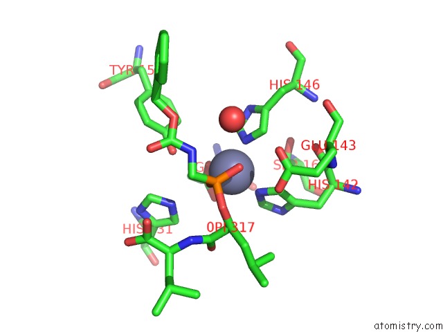
Mono view
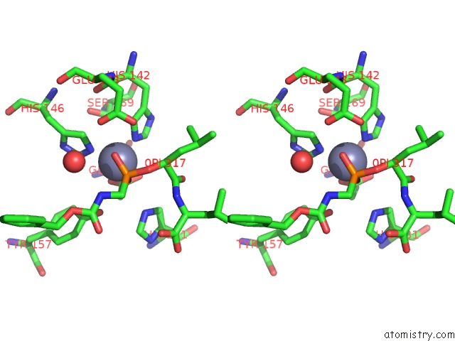
Stereo pair view
Mono view
Stereo pair view
A full contact list of Zinc with other atoms in the Zn binding
site number 1 of Structures of Two Thermolysin-Inhibitor Complexes That Differ By A Single Hydrogen Bond within 5.0Å range:
|
Reference:
D.E.Tronrud,
H.M.Holden,
B.W.Matthews.
Structures of Two Thermolysin-Inhibitor Complexes That Differ By A Single Hydrogen Bond. Science V. 235 571 1987.
ISSN: ISSN 0036-8075
PubMed: 3810156
Page generated: Tue Oct 29 08:07:22 2024
ISSN: ISSN 0036-8075
PubMed: 3810156
Last articles
Mg in 4JBTMg in 4JA9
Mg in 4JAS
Mg in 4JA7
Mg in 4J9V
Mg in 4JA2
Mg in 4J9Q
Mg in 4J9O
Mg in 4J9N
Mg in 4J9M