Zinc »
PDB 6jev-6jss »
6jn7 »
Zinc in PDB 6jn7: Structure of H216A Mutant Closed Form Peptidoglycan Peptidase
Protein crystallography data
The structure of Structure of H216A Mutant Closed Form Peptidoglycan Peptidase, PDB code: 6jn7
was solved by
K.J.Min,
D.R.An,
H.J.Yoon,
S.W.Suh,
H.H.Lee,
with X-Ray Crystallography technique. A brief refinement statistics is given in the table below:
| Resolution Low / High (Å) | 32.70 / 2.04 |
| Space group | P 1 21 1 |
| Cell size a, b, c (Å), α, β, γ (°) | 82.304, 105.632, 86.161, 90.00, 107.22, 90.00 |
| R / Rfree (%) | 16.7 / 19.4 |
Zinc Binding Sites:
The binding sites of Zinc atom in the Structure of H216A Mutant Closed Form Peptidoglycan Peptidase
(pdb code 6jn7). This binding sites where shown within
5.0 Angstroms radius around Zinc atom.
In total 3 binding sites of Zinc where determined in the Structure of H216A Mutant Closed Form Peptidoglycan Peptidase, PDB code: 6jn7:
Jump to Zinc binding site number: 1; 2; 3;
In total 3 binding sites of Zinc where determined in the Structure of H216A Mutant Closed Form Peptidoglycan Peptidase, PDB code: 6jn7:
Jump to Zinc binding site number: 1; 2; 3;
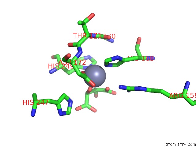
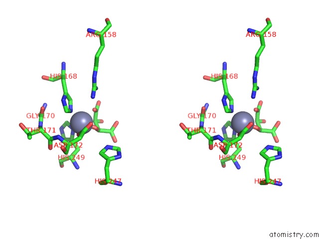
Zinc binding site 1 out of 3 in 6jn7
Go back to
Zinc binding site 1 out
of 3 in the Structure of H216A Mutant Closed Form Peptidoglycan Peptidase
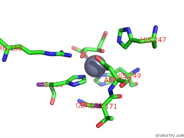
Mono view
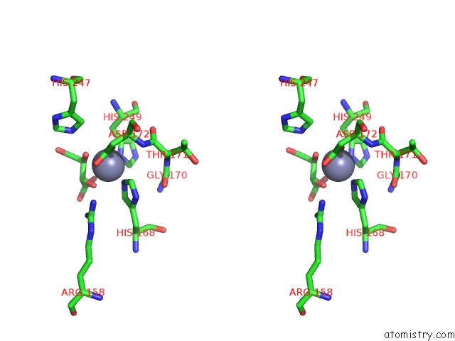
Stereo pair view
Mono view
Stereo pair view
A full contact list of Zinc with other atoms in the Zn binding
site number 1 of Structure of H216A Mutant Closed Form Peptidoglycan Peptidase within 5.0Å range:
|
Zinc binding site 2 out of 3 in 6jn7
Go back to
Zinc binding site 2 out
of 3 in the Structure of H216A Mutant Closed Form Peptidoglycan Peptidase
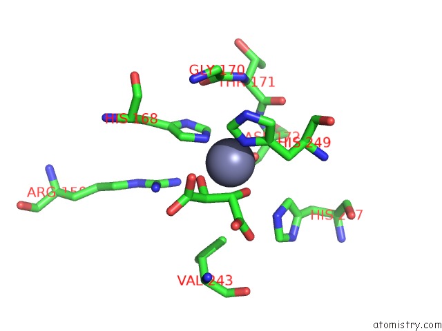
Mono view
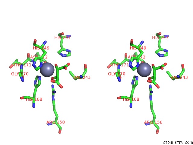
Stereo pair view
Mono view
Stereo pair view
A full contact list of Zinc with other atoms in the Zn binding
site number 2 of Structure of H216A Mutant Closed Form Peptidoglycan Peptidase within 5.0Å range:
|
Zinc binding site 3 out of 3 in 6jn7
Go back to
Zinc binding site 3 out
of 3 in the Structure of H216A Mutant Closed Form Peptidoglycan Peptidase
Mono view
Stereo pair view
Mono view
Stereo pair view
A full contact list of Zinc with other atoms in the Zn binding
site number 3 of Structure of H216A Mutant Closed Form Peptidoglycan Peptidase within 5.0Å range:
|
Reference:
S.Mobashery,
H.J.Yoon,
S.W.Suh,
H.H.Lee,
S.Ryu,
M.J.Lee,
D.Hesek,
K.J.Min,
D.R.An,
N.Rana,
S.J.Park,
B.M.Kim,
J.S.Kim.
Peptidoglycan Reshaping By A Noncanonical Peptidase For Helical Cell Shape in Campylobacter Jejuni Nat Commun 2020.
ISSN: ESSN 2041-1723
DOI: 10.1038/S41467-019-13934-4
Page generated: Thu Aug 21 16:27:20 2025
ISSN: ESSN 2041-1723
DOI: 10.1038/S41467-019-13934-4
Last articles
Zn in 6SI3Zn in 6SI2
Zn in 6SI1
Zn in 6SI0
Zn in 6SHZ
Zn in 6SHO
Zn in 6SHX
Zn in 6SHV
Zn in 6SHK
Zn in 6SHB