Zinc »
PDB 6jev-6jss »
6jew »
Zinc in PDB 6jew: K3U Bound Crystal Peptide Deformylase From Acinetobacter Baumanii
Enzymatic activity of K3U Bound Crystal Peptide Deformylase From Acinetobacter Baumanii
All present enzymatic activity of K3U Bound Crystal Peptide Deformylase From Acinetobacter Baumanii:
3.5.1.88;
3.5.1.88;
Protein crystallography data
The structure of K3U Bound Crystal Peptide Deformylase From Acinetobacter Baumanii, PDB code: 6jew
was solved by
I.H.Lee,
T.H.Ho,
L.W.Kang,
with X-Ray Crystallography technique. A brief refinement statistics is given in the table below:
| Resolution Low / High (Å) | 49.99 / 2.00 |
| Space group | P 32 |
| Cell size a, b, c (Å), α, β, γ (°) | 39.493, 39.493, 187.119, 90.00, 90.00, 120.00 |
| R / Rfree (%) | 22.6 / 29.2 |
Other elements in 6jew:
The structure of K3U Bound Crystal Peptide Deformylase From Acinetobacter Baumanii also contains other interesting chemical elements:
| Fluorine | (F) | 3 atoms |
Zinc Binding Sites:
The binding sites of Zinc atom in the K3U Bound Crystal Peptide Deformylase From Acinetobacter Baumanii
(pdb code 6jew). This binding sites where shown within
5.0 Angstroms radius around Zinc atom.
In total 2 binding sites of Zinc where determined in the K3U Bound Crystal Peptide Deformylase From Acinetobacter Baumanii, PDB code: 6jew:
Jump to Zinc binding site number: 1; 2;
In total 2 binding sites of Zinc where determined in the K3U Bound Crystal Peptide Deformylase From Acinetobacter Baumanii, PDB code: 6jew:
Jump to Zinc binding site number: 1; 2;
Zinc binding site 1 out of 2 in 6jew
Go back to
Zinc binding site 1 out
of 2 in the K3U Bound Crystal Peptide Deformylase From Acinetobacter Baumanii
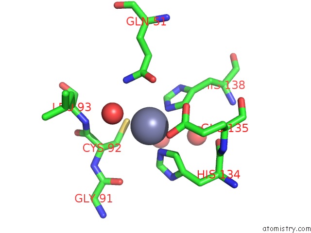
Mono view
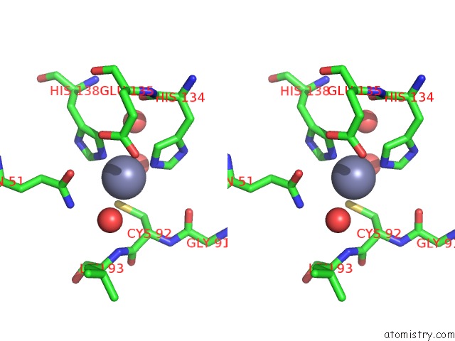
Stereo pair view
Mono view
Stereo pair view
A full contact list of Zinc with other atoms in the Zn binding
site number 1 of K3U Bound Crystal Peptide Deformylase From Acinetobacter Baumanii within 5.0Å range:
|
Zinc binding site 2 out of 2 in 6jew
Go back to
Zinc binding site 2 out
of 2 in the K3U Bound Crystal Peptide Deformylase From Acinetobacter Baumanii
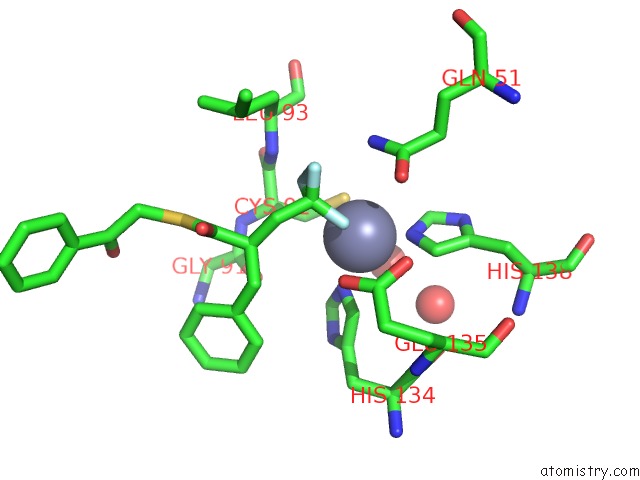
Mono view
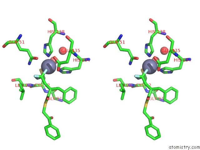
Stereo pair view
Mono view
Stereo pair view
A full contact list of Zinc with other atoms in the Zn binding
site number 2 of K3U Bound Crystal Peptide Deformylase From Acinetobacter Baumanii within 5.0Å range:
|
Reference:
I.H.Lee,
T.H.Ho,
L.W.Kang.
K3U Bound Crystal Peptide Deformylase From Acinetobacter Baumanii To Be Published.
Page generated: Thu Aug 21 16:22:08 2025
Last articles
Zn in 6SIXZn in 6SIW
Zn in 6SIV
Zn in 6SIC
Zn in 6SI4
Zn in 6SI3
Zn in 6SI2
Zn in 6SI1
Zn in 6SI0
Zn in 6SHZ