Zinc »
PDB 6j4n-6jeu »
6jdc »
Zinc in PDB 6jdc: Crystal Structure of N-Acetyl Mannosmaine Kinase in Complex with Mannac From Haemophilus Influenzae
Enzymatic activity of Crystal Structure of N-Acetyl Mannosmaine Kinase in Complex with Mannac From Haemophilus Influenzae
All present enzymatic activity of Crystal Structure of N-Acetyl Mannosmaine Kinase in Complex with Mannac From Haemophilus Influenzae:
2.7.1.60;
2.7.1.60;
Protein crystallography data
The structure of Crystal Structure of N-Acetyl Mannosmaine Kinase in Complex with Mannac From Haemophilus Influenzae, PDB code: 6jdc
was solved by
G.Thanuja,
S.Ramaswamy,
with X-Ray Crystallography technique. A brief refinement statistics is given in the table below:
| Resolution Low / High (Å) | 46.01 / 2.27 |
| Space group | I 41 2 2 |
| Cell size a, b, c (Å), α, β, γ (°) | 91.102, 91.102, 184.033, 90.00, 90.00, 90.00 |
| R / Rfree (%) | 21.7 / 26.6 |
Zinc Binding Sites:
The binding sites of Zinc atom in the Crystal Structure of N-Acetyl Mannosmaine Kinase in Complex with Mannac From Haemophilus Influenzae
(pdb code 6jdc). This binding sites where shown within
5.0 Angstroms radius around Zinc atom.
In total only one binding site of Zinc was determined in the Crystal Structure of N-Acetyl Mannosmaine Kinase in Complex with Mannac From Haemophilus Influenzae, PDB code: 6jdc:
In total only one binding site of Zinc was determined in the Crystal Structure of N-Acetyl Mannosmaine Kinase in Complex with Mannac From Haemophilus Influenzae, PDB code: 6jdc:
Zinc binding site 1 out of 1 in 6jdc
Go back to
Zinc binding site 1 out
of 1 in the Crystal Structure of N-Acetyl Mannosmaine Kinase in Complex with Mannac From Haemophilus Influenzae
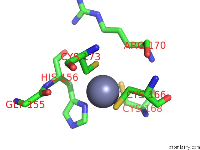
Mono view
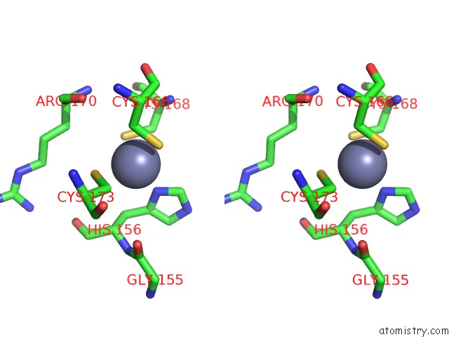
Stereo pair view
Mono view
Stereo pair view
A full contact list of Zinc with other atoms in the Zn binding
site number 1 of Crystal Structure of N-Acetyl Mannosmaine Kinase in Complex with Mannac From Haemophilus Influenzae within 5.0Å range:
|
Reference:
G.Thanuja,
S.Ramaswamy.
Crystal Structure of N-Acetyl Mannosmaine Kinase in Complex with Mannac From Haemophilus Influenzae To Be Published.
Page generated: Thu Aug 21 16:20:12 2025
Last articles
Zn in 6S6YZn in 6S53
Zn in 6S7F
Zn in 6S6B
Zn in 6S3V
Zn in 6S3J
Zn in 6S3A
Zn in 6S3G
Zn in 6S1X
Zn in 6S2B