Zinc »
PDB 6rwo-6s6b »
6s3a »
Zinc in PDB 6s3a: Coxsackie B3 2C Protein in Complex with S-Fluoxetine
Protein crystallography data
The structure of Coxsackie B3 2C Protein in Complex with S-Fluoxetine, PDB code: 6s3a
was solved by
P.El Kazzi,
N.Papageorgiou,
F.P.Ferron,
L.Bauer,
F.Van Kuppeveld,
B.Coutard,
with X-Ray Crystallography technique. A brief refinement statistics is given in the table below:
| Resolution Low / High (Å) | 44.14 / 1.52 |
| Space group | P 21 21 21 |
| Cell size a, b, c (Å), α, β, γ (°) | 48.398, 53.183, 79.152, 90, 90, 90 |
| R / Rfree (%) | n/a / 20 |
Other elements in 6s3a:
The structure of Coxsackie B3 2C Protein in Complex with S-Fluoxetine also contains other interesting chemical elements:
| Chlorine | (Cl) | 1 atom |
| Fluorine | (F) | 3 atoms |
Zinc Binding Sites:
The binding sites of Zinc atom in the Coxsackie B3 2C Protein in Complex with S-Fluoxetine
(pdb code 6s3a). This binding sites where shown within
5.0 Angstroms radius around Zinc atom.
In total only one binding site of Zinc was determined in the Coxsackie B3 2C Protein in Complex with S-Fluoxetine, PDB code: 6s3a:
In total only one binding site of Zinc was determined in the Coxsackie B3 2C Protein in Complex with S-Fluoxetine, PDB code: 6s3a:
Zinc binding site 1 out of 1 in 6s3a
Go back to
Zinc binding site 1 out
of 1 in the Coxsackie B3 2C Protein in Complex with S-Fluoxetine
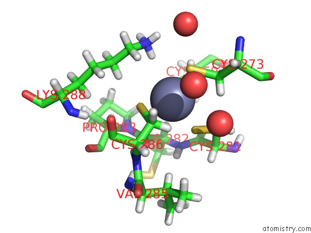
Mono view
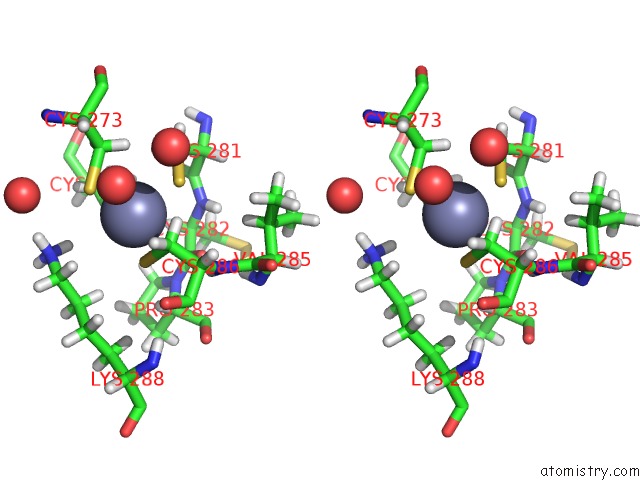
Stereo pair view
Mono view
Stereo pair view
A full contact list of Zinc with other atoms in the Zn binding
site number 1 of Coxsackie B3 2C Protein in Complex with S-Fluoxetine within 5.0Å range:
|
Reference:
P.El Kazzi,
L.Bauer,
N.Papageorgiou,
F.P.Ferron,
E.Decroly,
B.Canard,
A.Brancale,
F.Van Kupperveld,
B.Coutard.
The Crystal Structure of Enterovirus 2C Catalytic Domain in Complex Withs-Fluoxetine Reveals Its Antiviral Mode of Action. To Be Published.
Page generated: Tue Oct 29 07:01:14 2024
Last articles
Mg in 1S8FMg in 1S83
Mg in 1S77
Mg in 1S76
Mg in 1S4E
Mg in 1S6P
Mg in 1S6H
Mg in 1S5J
Mg in 1S5G
Mg in 1S2V