Zinc »
PDB 5vem-5vmz »
5vge »
Zinc in PDB 5vge: Crystal Structure of Hla-C*07:02 in Complex with Ryr Peptide
Protein crystallography data
The structure of Crystal Structure of Hla-C*07:02 in Complex with Ryr Peptide, PDB code: 5vge
was solved by
J.I.Mobbs,
J.P.Vivian,
S.Gras,
J.Rossjohn,
with X-Ray Crystallography technique. A brief refinement statistics is given in the table below:
| Resolution Low / High (Å) | 43.16 / 2.60 |
| Space group | P 65 |
| Cell size a, b, c (Å), α, β, γ (°) | 99.670, 99.670, 77.914, 90.00, 90.00, 120.00 |
| R / Rfree (%) | 16.3 / 22.1 |
Zinc Binding Sites:
The binding sites of Zinc atom in the Crystal Structure of Hla-C*07:02 in Complex with Ryr Peptide
(pdb code 5vge). This binding sites where shown within
5.0 Angstroms radius around Zinc atom.
In total 3 binding sites of Zinc where determined in the Crystal Structure of Hla-C*07:02 in Complex with Ryr Peptide, PDB code: 5vge:
Jump to Zinc binding site number: 1; 2; 3;
In total 3 binding sites of Zinc where determined in the Crystal Structure of Hla-C*07:02 in Complex with Ryr Peptide, PDB code: 5vge:
Jump to Zinc binding site number: 1; 2; 3;
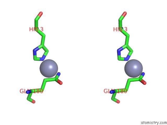
Zinc binding site 1 out of 3 in 5vge
Go back to
Zinc binding site 1 out
of 3 in the Crystal Structure of Hla-C*07:02 in Complex with Ryr Peptide

Mono view
Stereo pair view
Mono view
Stereo pair view
A full contact list of Zinc with other atoms in the Zn binding
site number 1 of Crystal Structure of Hla-C*07:02 in Complex with Ryr Peptide within 5.0Å range:
|
Zinc binding site 2 out of 3 in 5vge
Go back to
Zinc binding site 2 out
of 3 in the Crystal Structure of Hla-C*07:02 in Complex with Ryr Peptide

Mono view
Stereo pair view
Mono view
Stereo pair view
A full contact list of Zinc with other atoms in the Zn binding
site number 2 of Crystal Structure of Hla-C*07:02 in Complex with Ryr Peptide within 5.0Å range:
|
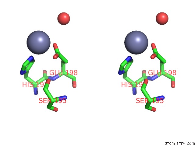
Zinc binding site 3 out of 3 in 5vge
Go back to
Zinc binding site 3 out
of 3 in the Crystal Structure of Hla-C*07:02 in Complex with Ryr Peptide

Mono view

Stereo pair view
Mono view
Stereo pair view
A full contact list of Zinc with other atoms in the Zn binding
site number 3 of Crystal Structure of Hla-C*07:02 in Complex with Ryr Peptide within 5.0Å range:
|
Reference:
G.Kaur,
S.Gras,
J.I.Mobbs,
J.P.Vivian,
A.Cortes,
T.Barber,
S.B.Kuttikkatte,
L.T.Jensen,
K.E.Attfield,
C.A.Dendrou,
M.Carrington,
G.Mcvean,
A.W.Purcell,
J.Rossjohn,
L.Fugger.
Structural and Regulatory Diversity Shape Hla-C Protein Expression Levels. Nat Commun V. 8 15924 2017.
ISSN: ESSN 2041-1723
PubMed: 28649982
DOI: 10.1038/NCOMMS15924
Page generated: Thu Aug 21 09:57:33 2025
ISSN: ESSN 2041-1723
PubMed: 28649982
DOI: 10.1038/NCOMMS15924
Last articles
Zn in 6FF7Zn in 6FF4
Zn in 6FFZ
Zn in 6FFW
Zn in 6FF9
Zn in 6FET
Zn in 6FE7
Zn in 6FEB
Zn in 6FEA
Zn in 6FE6