Zinc »
PDB 6f9r-6fhg »
6ffz »
Zinc in PDB 6ffz: Crystal Structure Analysis of the Clock Protein EA4 (Glycosylation Form)
Protein crystallography data
The structure of Crystal Structure Analysis of the Clock Protein EA4 (Glycosylation Form), PDB code: 6ffz
was solved by
S.-Y.Park,
T.Hiraki,
with X-Ray Crystallography technique. A brief refinement statistics is given in the table below:
| Resolution Low / High (Å) | 20.00 / 2.11 |
| Space group | P 1 21 1 |
| Cell size a, b, c (Å), α, β, γ (°) | 47.098, 73.894, 47.446, 90.00, 104.07, 90.00 |
| R / Rfree (%) | 17 / 25.1 |
Zinc Binding Sites:
The binding sites of Zinc atom in the Crystal Structure Analysis of the Clock Protein EA4 (Glycosylation Form)
(pdb code 6ffz). This binding sites where shown within
5.0 Angstroms radius around Zinc atom.
In total 2 binding sites of Zinc where determined in the Crystal Structure Analysis of the Clock Protein EA4 (Glycosylation Form), PDB code: 6ffz:
Jump to Zinc binding site number: 1; 2;
In total 2 binding sites of Zinc where determined in the Crystal Structure Analysis of the Clock Protein EA4 (Glycosylation Form), PDB code: 6ffz:
Jump to Zinc binding site number: 1; 2;
Zinc binding site 1 out of 2 in 6ffz
Go back to
Zinc binding site 1 out
of 2 in the Crystal Structure Analysis of the Clock Protein EA4 (Glycosylation Form)
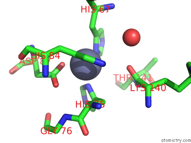
Mono view
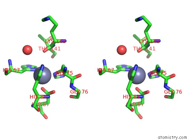
Stereo pair view
Mono view
Stereo pair view
A full contact list of Zinc with other atoms in the Zn binding
site number 1 of Crystal Structure Analysis of the Clock Protein EA4 (Glycosylation Form) within 5.0Å range:
|
Zinc binding site 2 out of 2 in 6ffz
Go back to
Zinc binding site 2 out
of 2 in the Crystal Structure Analysis of the Clock Protein EA4 (Glycosylation Form)
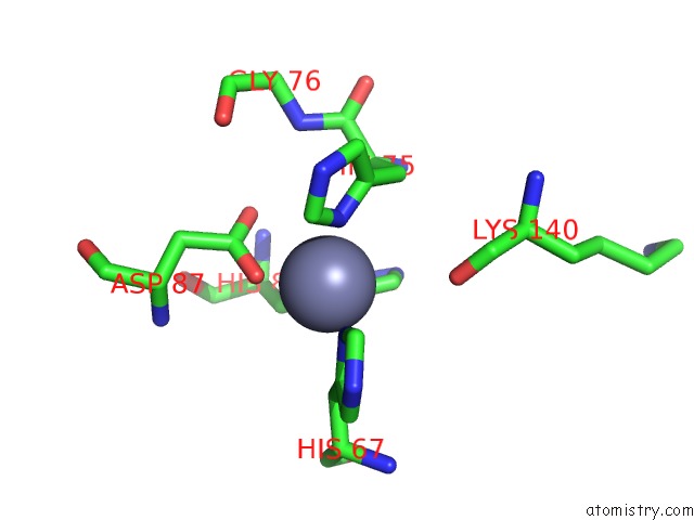
Mono view
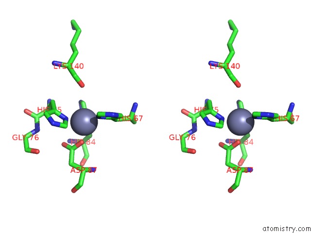
Stereo pair view
Mono view
Stereo pair view
A full contact list of Zinc with other atoms in the Zn binding
site number 2 of Crystal Structure Analysis of the Clock Protein EA4 (Glycosylation Form) within 5.0Å range:
|
Reference:
T.Hiraki,
N.Shibayama,
J.R.M.Tame,
S.Akashi,
S.-Y.Park.
The Clock Protein EA4 Ticks Away with Movement of A Mobile Copper Ion To Be Published.
Page generated: Mon Oct 28 20:57:20 2024
Last articles
Fe in 3OO3Fe in 3OMI
Fe in 3OM3
Fe in 3OMA
Fe in 3OFU
Fe in 3OJT
Fe in 3OK5
Fe in 3OL5
Fe in 3OJ1
Fe in 3OJG