Zinc »
PDB 5o2f-5ofb »
5od6 »
Zinc in PDB 5od6: Crystal Structure of SMAD3-MH1 Bound to the Ggcgc Site.
Protein crystallography data
The structure of Crystal Structure of SMAD3-MH1 Bound to the Ggcgc Site., PDB code: 5od6
was solved by
Z.Kaczmarska,
J.A.Marquez,
M.J.Macias,
with X-Ray Crystallography technique. A brief refinement statistics is given in the table below:
| Resolution Low / High (Å) | 27.11 / 2.00 |
| Space group | I 41 |
| Cell size a, b, c (Å), α, β, γ (°) | 105.200, 105.200, 73.240, 90.00, 90.00, 90.00 |
| R / Rfree (%) | 19.5 / 23.5 |
Zinc Binding Sites:
The binding sites of Zinc atom in the Crystal Structure of SMAD3-MH1 Bound to the Ggcgc Site.
(pdb code 5od6). This binding sites where shown within
5.0 Angstroms radius around Zinc atom.
In total 2 binding sites of Zinc where determined in the Crystal Structure of SMAD3-MH1 Bound to the Ggcgc Site., PDB code: 5od6:
Jump to Zinc binding site number: 1; 2;
In total 2 binding sites of Zinc where determined in the Crystal Structure of SMAD3-MH1 Bound to the Ggcgc Site., PDB code: 5od6:
Jump to Zinc binding site number: 1; 2;
Zinc binding site 1 out of 2 in 5od6
Go back to
Zinc binding site 1 out
of 2 in the Crystal Structure of SMAD3-MH1 Bound to the Ggcgc Site.
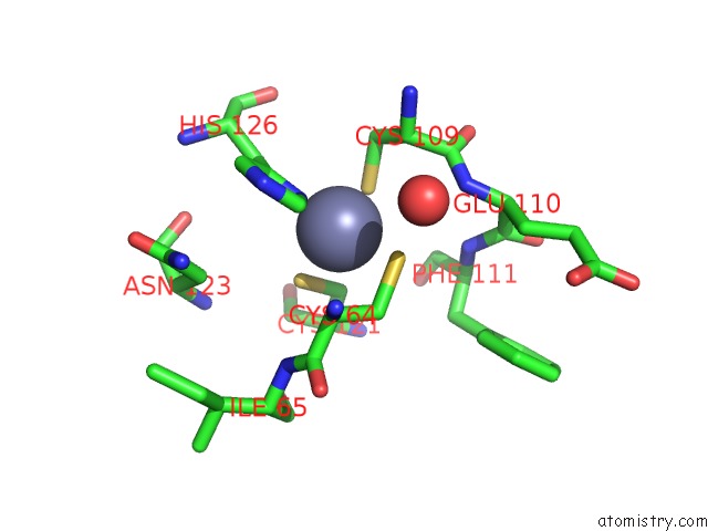
Mono view
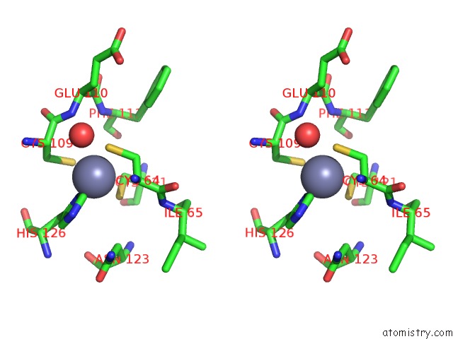
Stereo pair view
Mono view
Stereo pair view
A full contact list of Zinc with other atoms in the Zn binding
site number 1 of Crystal Structure of SMAD3-MH1 Bound to the Ggcgc Site. within 5.0Å range:
|
Zinc binding site 2 out of 2 in 5od6
Go back to
Zinc binding site 2 out
of 2 in the Crystal Structure of SMAD3-MH1 Bound to the Ggcgc Site.
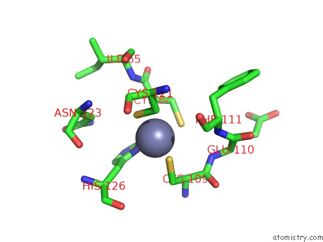
Mono view
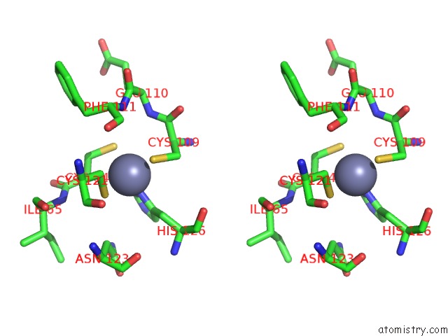
Stereo pair view
Mono view
Stereo pair view
A full contact list of Zinc with other atoms in the Zn binding
site number 2 of Crystal Structure of SMAD3-MH1 Bound to the Ggcgc Site. within 5.0Å range:
|
Reference:
P.Martin-Malpartida,
M.Batet,
Z.Kaczmarska,
R.Freier,
T.Gomes,
E.Aragon,
Y.Zou,
Q.Wang,
Q.Xi,
L.Ruiz,
A.Vea,
J.A.Marquez,
J.Massague,
M.J.Macias.
Structural Basis For Genome Wide Recognition of 5-Bp Gc Motifs By Smad Transcription Factors. Nat Commun V. 8 2070 2017.
ISSN: ESSN 2041-1723
PubMed: 29234012
DOI: 10.1038/S41467-017-02054-6
Page generated: Sun Oct 27 23:25:37 2024
ISSN: ESSN 2041-1723
PubMed: 29234012
DOI: 10.1038/S41467-017-02054-6
Last articles
Mg in 1VR0Mg in 1VQM
Mg in 1VQL
Mg in 1VQK
Mg in 1VQ9
Mg in 1VQ7
Mg in 1VQ5
Mg in 1VQ6
Mg in 1VQ8
Mg in 1VQ4