Zinc »
PDB 5i3p-5ikf »
5ib9 »
Zinc in PDB 5ib9: Crystal Structure of Aminopeptidase Equipped with Pad From Aneurinibacillus Sp. Am-1
Protein crystallography data
The structure of Crystal Structure of Aminopeptidase Equipped with Pad From Aneurinibacillus Sp. Am-1, PDB code: 5ib9
was solved by
R.Tagawa,
H.Nakano,
K.Watanabe,
with X-Ray Crystallography technique. A brief refinement statistics is given in the table below:
| Resolution Low / High (Å) | 39.80 / 1.40 |
| Space group | P 21 21 2 |
| Cell size a, b, c (Å), α, β, γ (°) | 93.180, 68.443, 76.589, 90.00, 90.00, 90.00 |
| R / Rfree (%) | 17.9 / 22.3 |
Zinc Binding Sites:
The binding sites of Zinc atom in the Crystal Structure of Aminopeptidase Equipped with Pad From Aneurinibacillus Sp. Am-1
(pdb code 5ib9). This binding sites where shown within
5.0 Angstroms radius around Zinc atom.
In total 6 binding sites of Zinc where determined in the Crystal Structure of Aminopeptidase Equipped with Pad From Aneurinibacillus Sp. Am-1, PDB code: 5ib9:
Jump to Zinc binding site number: 1; 2; 3; 4; 5; 6;
In total 6 binding sites of Zinc where determined in the Crystal Structure of Aminopeptidase Equipped with Pad From Aneurinibacillus Sp. Am-1, PDB code: 5ib9:
Jump to Zinc binding site number: 1; 2; 3; 4; 5; 6;
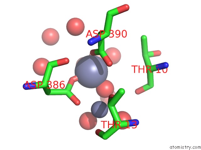
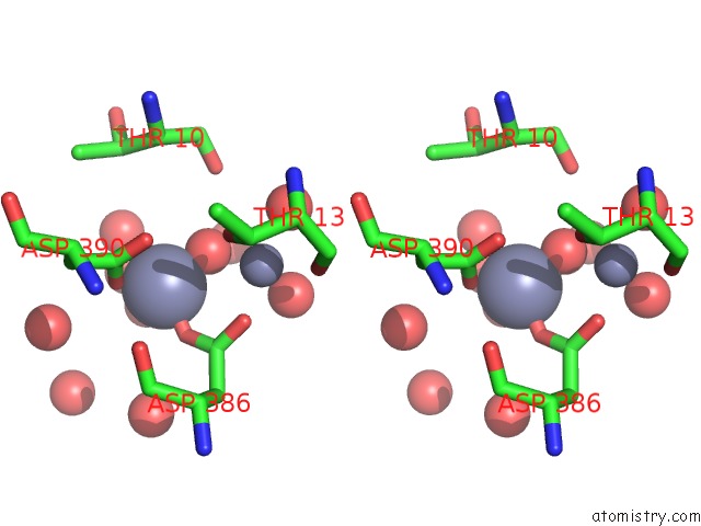
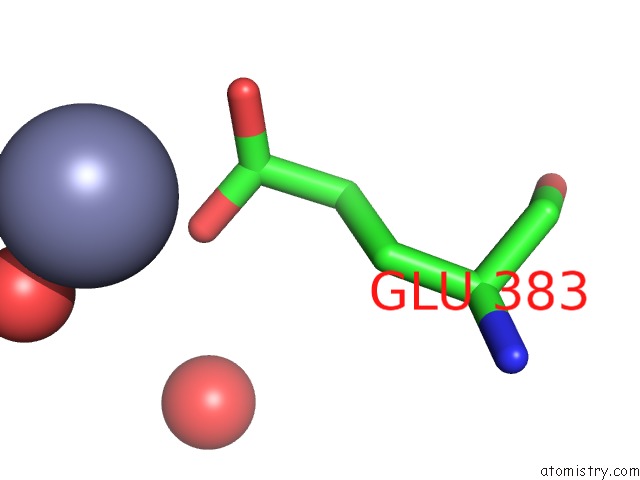
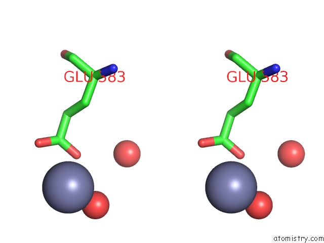
Zinc binding site 1 out of 6 in 5ib9
Go back to
Zinc binding site 1 out
of 6 in the Crystal Structure of Aminopeptidase Equipped with Pad From Aneurinibacillus Sp. Am-1
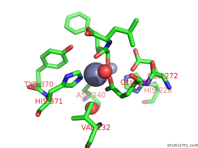
Mono view
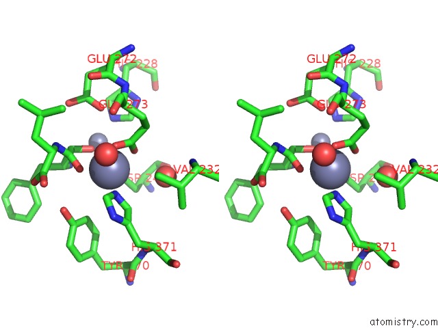
Stereo pair view
Mono view
Stereo pair view
A full contact list of Zinc with other atoms in the Zn binding
site number 1 of Crystal Structure of Aminopeptidase Equipped with Pad From Aneurinibacillus Sp. Am-1 within 5.0Å range:
|
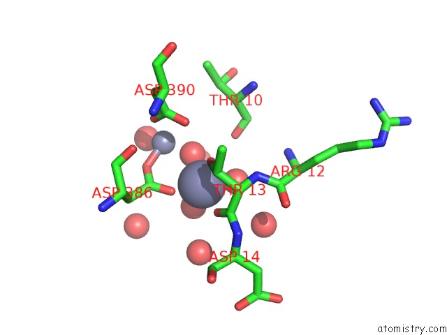
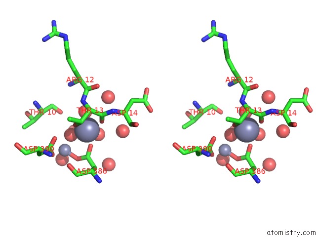
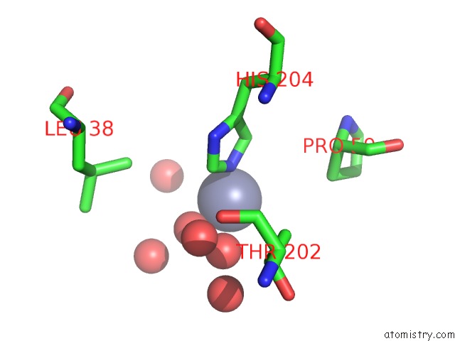
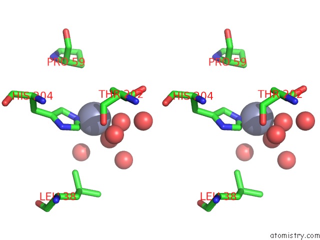
Zinc binding site 2 out of 6 in 5ib9
Go back to
Zinc binding site 2 out
of 6 in the Crystal Structure of Aminopeptidase Equipped with Pad From Aneurinibacillus Sp. Am-1
Mono view
Stereo pair view
Mono view
Stereo pair view
A full contact list of Zinc with other atoms in the Zn binding
site number 2 of Crystal Structure of Aminopeptidase Equipped with Pad From Aneurinibacillus Sp. Am-1 within 5.0Å range:
|
Zinc binding site 3 out of 6 in 5ib9
Go back to
Zinc binding site 3 out
of 6 in the Crystal Structure of Aminopeptidase Equipped with Pad From Aneurinibacillus Sp. Am-1
Mono view
Stereo pair view
Mono view
Stereo pair view
A full contact list of Zinc with other atoms in the Zn binding
site number 3 of Crystal Structure of Aminopeptidase Equipped with Pad From Aneurinibacillus Sp. Am-1 within 5.0Å range:
|
Zinc binding site 4 out of 6 in 5ib9
Go back to
Zinc binding site 4 out
of 6 in the Crystal Structure of Aminopeptidase Equipped with Pad From Aneurinibacillus Sp. Am-1
Mono view
Stereo pair view
Mono view
Stereo pair view
A full contact list of Zinc with other atoms in the Zn binding
site number 4 of Crystal Structure of Aminopeptidase Equipped with Pad From Aneurinibacillus Sp. Am-1 within 5.0Å range:
|
Zinc binding site 5 out of 6 in 5ib9
Go back to
Zinc binding site 5 out
of 6 in the Crystal Structure of Aminopeptidase Equipped with Pad From Aneurinibacillus Sp. Am-1
Mono view
Stereo pair view
Mono view
Stereo pair view
A full contact list of Zinc with other atoms in the Zn binding
site number 5 of Crystal Structure of Aminopeptidase Equipped with Pad From Aneurinibacillus Sp. Am-1 within 5.0Å range:
|
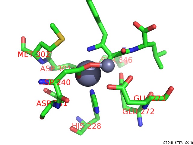
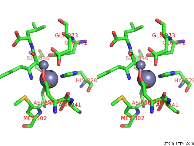
Zinc binding site 6 out of 6 in 5ib9
Go back to
Zinc binding site 6 out
of 6 in the Crystal Structure of Aminopeptidase Equipped with Pad From Aneurinibacillus Sp. Am-1
Mono view
Stereo pair view
Mono view
Stereo pair view
A full contact list of Zinc with other atoms in the Zn binding
site number 6 of Crystal Structure of Aminopeptidase Equipped with Pad From Aneurinibacillus Sp. Am-1 within 5.0Å range:
|
Reference:
R.Tagawa,
H.Nakano,
K.Watanabe.
Crystal Structure of Aminopeptidase To Be Published.
Page generated: Thu Aug 21 03:23:24 2025
Last articles
Zn in 5SE0Zn in 5SDZ
Zn in 5SDX
Zn in 5SE1
Zn in 5SDY
Zn in 5SDW
Zn in 5ROB
Zn in 5SDV
Zn in 5SDU
Zn in 5SBG