Zinc »
PDB 5brv-5c3k »
5c11 »
Zinc in PDB 5c11: Crystal Structure of JARID1A Phd Finger Bound to Histone H3C4ME3 Peptide
Protein crystallography data
The structure of Crystal Structure of JARID1A Phd Finger Bound to Histone H3C4ME3 Peptide, PDB code: 5c11
was solved by
J.Huang,
H.Li,
with X-Ray Crystallography technique. A brief refinement statistics is given in the table below:
| Resolution Low / High (Å) | 44.46 / 2.80 |
| Space group | I 4 3 2 |
| Cell size a, b, c (Å), α, β, γ (°) | 108.910, 108.910, 108.910, 90.00, 90.00, 90.00 |
| R / Rfree (%) | 24.6 / 27.9 |
Zinc Binding Sites:
The binding sites of Zinc atom in the Crystal Structure of JARID1A Phd Finger Bound to Histone H3C4ME3 Peptide
(pdb code 5c11). This binding sites where shown within
5.0 Angstroms radius around Zinc atom.
In total 2 binding sites of Zinc where determined in the Crystal Structure of JARID1A Phd Finger Bound to Histone H3C4ME3 Peptide, PDB code: 5c11:
Jump to Zinc binding site number: 1; 2;
In total 2 binding sites of Zinc where determined in the Crystal Structure of JARID1A Phd Finger Bound to Histone H3C4ME3 Peptide, PDB code: 5c11:
Jump to Zinc binding site number: 1; 2;
Zinc binding site 1 out of 2 in 5c11
Go back to
Zinc binding site 1 out
of 2 in the Crystal Structure of JARID1A Phd Finger Bound to Histone H3C4ME3 Peptide
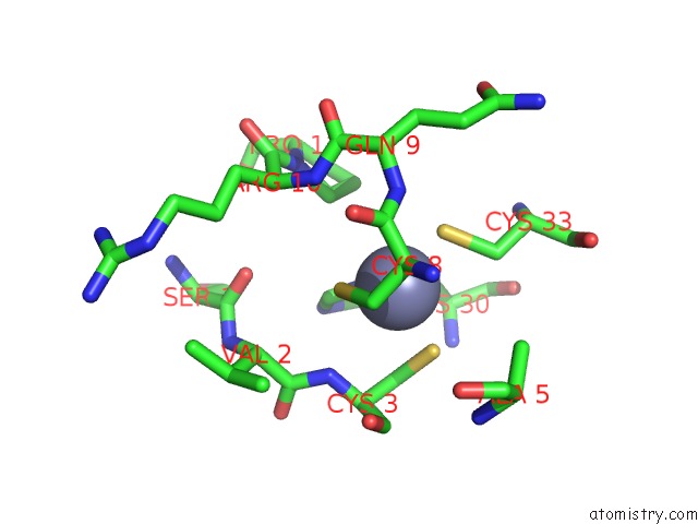
Mono view
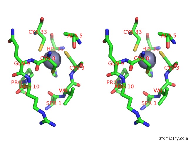
Stereo pair view
Mono view
Stereo pair view
A full contact list of Zinc with other atoms in the Zn binding
site number 1 of Crystal Structure of JARID1A Phd Finger Bound to Histone H3C4ME3 Peptide within 5.0Å range:
|
Zinc binding site 2 out of 2 in 5c11
Go back to
Zinc binding site 2 out
of 2 in the Crystal Structure of JARID1A Phd Finger Bound to Histone H3C4ME3 Peptide
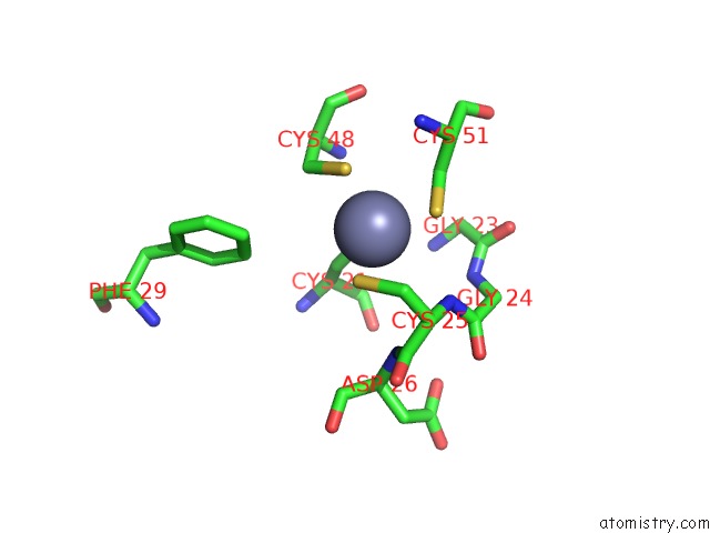
Mono view
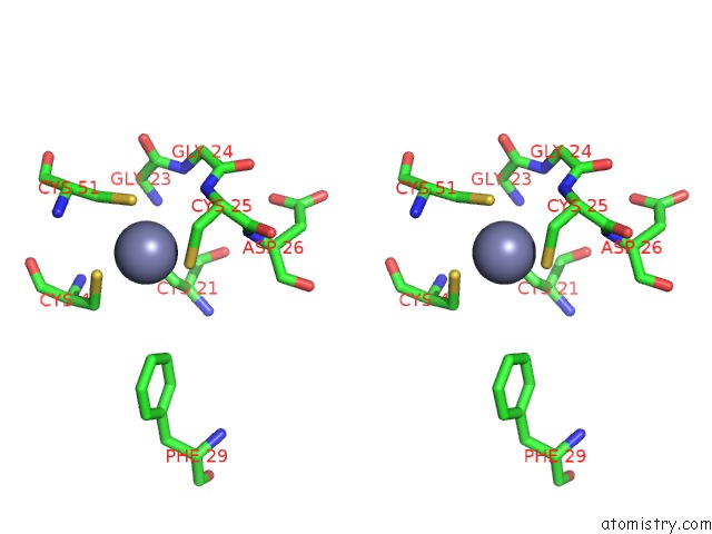
Stereo pair view
Mono view
Stereo pair view
A full contact list of Zinc with other atoms in the Zn binding
site number 2 of Crystal Structure of JARID1A Phd Finger Bound to Histone H3C4ME3 Peptide within 5.0Å range:
|
Reference:
J.Huang,
H.Li.
Crystal Structure of JARID1A Phd Finger Bound to Histone H3C4ME3 Peptide Nat Commun 2015.
ISSN: ESSN 2041-1723
Page generated: Sun Oct 27 13:38:39 2024
ISSN: ESSN 2041-1723
Last articles
Na in 8W46Na in 8W45
Na in 8W44
Na in 8W1Y
Na in 8W43
Na in 8W1W
Na in 8W42
Na in 8VU4
Na in 8W2C
Na in 8W1X