Zinc »
PDB 4y93-4ymk »
4yg2 »
Zinc in PDB 4yg2: X-Ray Crystal Structur of Escherichia Coli Rna Polymerase SIGMA70 Holoenzyme
Enzymatic activity of X-Ray Crystal Structur of Escherichia Coli Rna Polymerase SIGMA70 Holoenzyme
All present enzymatic activity of X-Ray Crystal Structur of Escherichia Coli Rna Polymerase SIGMA70 Holoenzyme:
2.7.7.6;
2.7.7.6;
Protein crystallography data
The structure of X-Ray Crystal Structur of Escherichia Coli Rna Polymerase SIGMA70 Holoenzyme, PDB code: 4yg2
was solved by
K.S.Murakami,
with X-Ray Crystallography technique. A brief refinement statistics is given in the table below:
| Resolution Low / High (Å) | 29.98 / 3.70 |
| Space group | P 21 21 21 |
| Cell size a, b, c (Å), α, β, γ (°) | 187.308, 205.901, 309.185, 90.00, 90.00, 90.00 |
| R / Rfree (%) | 19.1 / 26 |
Other elements in 4yg2:
The structure of X-Ray Crystal Structur of Escherichia Coli Rna Polymerase SIGMA70 Holoenzyme also contains other interesting chemical elements:
| Magnesium | (Mg) | 4 atoms |
Zinc Binding Sites:
The binding sites of Zinc atom in the X-Ray Crystal Structur of Escherichia Coli Rna Polymerase SIGMA70 Holoenzyme
(pdb code 4yg2). This binding sites where shown within
5.0 Angstroms radius around Zinc atom.
In total 4 binding sites of Zinc where determined in the X-Ray Crystal Structur of Escherichia Coli Rna Polymerase SIGMA70 Holoenzyme, PDB code: 4yg2:
Jump to Zinc binding site number: 1; 2; 3; 4;
In total 4 binding sites of Zinc where determined in the X-Ray Crystal Structur of Escherichia Coli Rna Polymerase SIGMA70 Holoenzyme, PDB code: 4yg2:
Jump to Zinc binding site number: 1; 2; 3; 4;
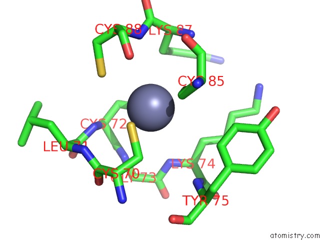
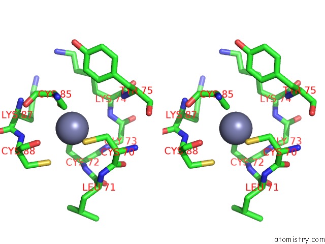
Zinc binding site 1 out of 4 in 4yg2
Go back to
Zinc binding site 1 out
of 4 in the X-Ray Crystal Structur of Escherichia Coli Rna Polymerase SIGMA70 Holoenzyme
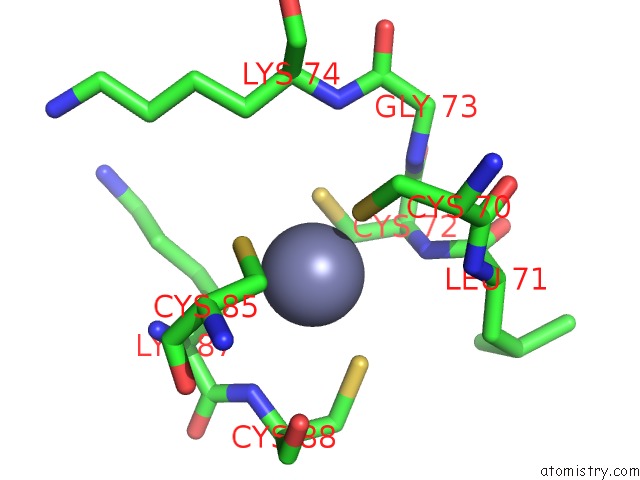
Mono view
Stereo pair view
Mono view
Stereo pair view
A full contact list of Zinc with other atoms in the Zn binding
site number 1 of X-Ray Crystal Structur of Escherichia Coli Rna Polymerase SIGMA70 Holoenzyme within 5.0Å range:
|
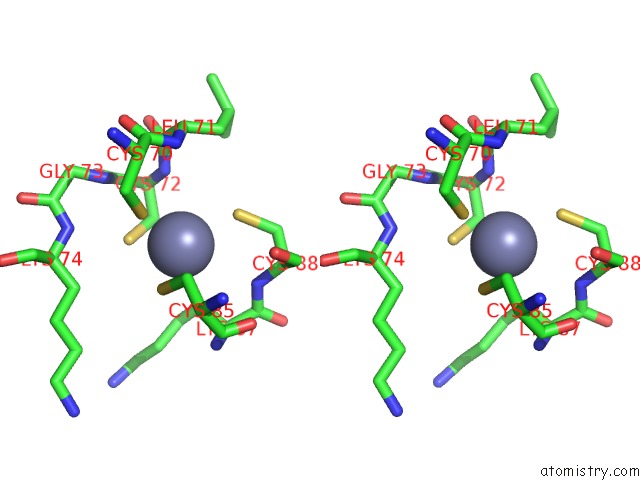
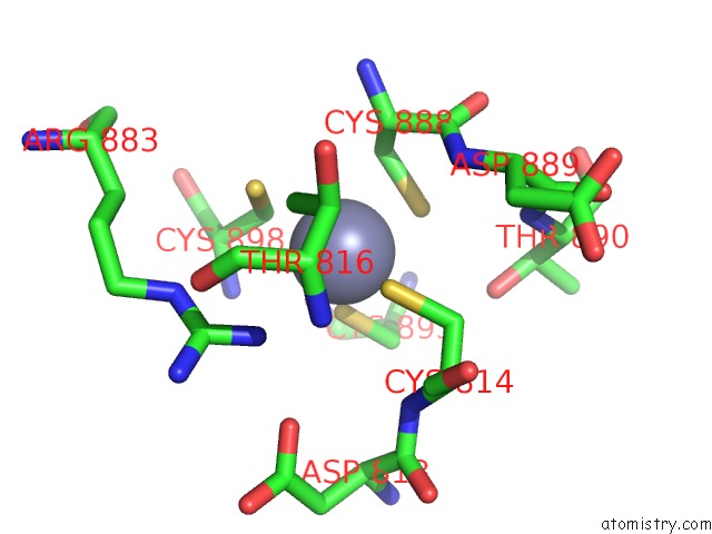
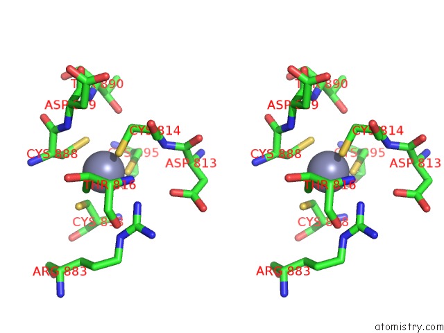
Zinc binding site 2 out of 4 in 4yg2
Go back to
Zinc binding site 2 out
of 4 in the X-Ray Crystal Structur of Escherichia Coli Rna Polymerase SIGMA70 Holoenzyme
Mono view
Stereo pair view
Mono view
Stereo pair view
A full contact list of Zinc with other atoms in the Zn binding
site number 2 of X-Ray Crystal Structur of Escherichia Coli Rna Polymerase SIGMA70 Holoenzyme within 5.0Å range:
|
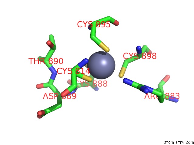
Zinc binding site 3 out of 4 in 4yg2
Go back to
Zinc binding site 3 out
of 4 in the X-Ray Crystal Structur of Escherichia Coli Rna Polymerase SIGMA70 Holoenzyme
Mono view
Stereo pair view
Mono view
Stereo pair view
A full contact list of Zinc with other atoms in the Zn binding
site number 3 of X-Ray Crystal Structur of Escherichia Coli Rna Polymerase SIGMA70 Holoenzyme within 5.0Å range:
|
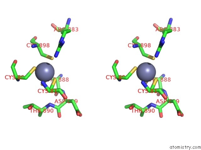
Zinc binding site 4 out of 4 in 4yg2
Go back to
Zinc binding site 4 out
of 4 in the X-Ray Crystal Structur of Escherichia Coli Rna Polymerase SIGMA70 Holoenzyme
Mono view
Stereo pair view
Mono view
Stereo pair view
A full contact list of Zinc with other atoms in the Zn binding
site number 4 of X-Ray Crystal Structur of Escherichia Coli Rna Polymerase SIGMA70 Holoenzyme within 5.0Å range:
|
Reference:
K.S.Murakami,
K.S.Murakami.
N/A N/A.
ISSN: ESSN 1083-351X
PubMed: 23389035
DOI: 10.1074/JBC.M112.430900
Page generated: Sun Oct 27 11:11:42 2024
ISSN: ESSN 1083-351X
PubMed: 23389035
DOI: 10.1074/JBC.M112.430900
Last articles
Mg in 4UM5Mg in 4UKD
Mg in 4UJ3
Mg in 4UJ5
Mg in 4UJ4
Mg in 4UHS
Mg in 4UHO
Mg in 4UHN
Mg in 4UHM
Mg in 4UHK