Zinc »
PDB 4w9y-4wnv »
4whb »
Zinc in PDB 4whb: Crystal Structure of Phenylurea Hydrolase B
Protein crystallography data
The structure of Crystal Structure of Phenylurea Hydrolase B, PDB code: 4whb
was solved by
E.Sugrue,
P.D.Carr,
J.L.Khurana,
C.J.Jackson,
with X-Ray Crystallography technique. A brief refinement statistics is given in the table below:
| Resolution Low / High (Å) | 39.60 / 2.96 |
| Space group | P 1 21 1 |
| Cell size a, b, c (Å), α, β, γ (°) | 77.374, 100.526, 238.551, 90.00, 98.37, 90.00 |
| R / Rfree (%) | 25.2 / 30.9 |
Zinc Binding Sites:
The binding sites of Zinc atom in the Crystal Structure of Phenylurea Hydrolase B
(pdb code 4whb). This binding sites where shown within
5.0 Angstroms radius around Zinc atom.
In total only one binding site of Zinc was determined in the Crystal Structure of Phenylurea Hydrolase B, PDB code: 4whb:
In total only one binding site of Zinc was determined in the Crystal Structure of Phenylurea Hydrolase B, PDB code: 4whb:
Zinc binding site 1 out of 1 in 4whb
Go back to
Zinc binding site 1 out
of 1 in the Crystal Structure of Phenylurea Hydrolase B
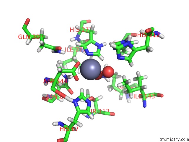
Mono view
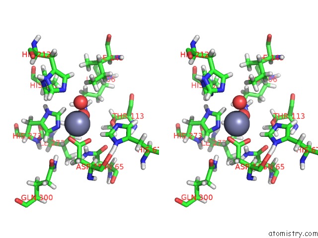
Stereo pair view
Mono view
Stereo pair view
A full contact list of Zinc with other atoms in the Zn binding
site number 1 of Crystal Structure of Phenylurea Hydrolase B within 5.0Å range:
|
Reference:
E.Sugrue,
N.J.Fraser,
D.H.Hopkins,
P.D.Carr,
J.L.Khurana,
J.G.Oakeshott,
C.Scott,
C.J.Jackson.
Evolutionary Expansion of the Amidohydrolase Superfamily in Response to Synthetic Compounds: Molinate and Diuron Hydrolases. To Be Published.
Page generated: Wed Aug 20 23:05:26 2025
Last articles
Zn in 5LUFZn in 5LTZ
Zn in 5LT8
Zn in 5LT7
Zn in 5LS6
Zn in 5LSV
Zn in 5LSZ
Zn in 5LSY
Zn in 5LSU
Zn in 5LSX