Zinc »
PDB 4qim-4qsa »
4qoe »
Zinc in PDB 4qoe: The Value 'Crystal Structure of Fad Quinone Reductase 2 at 1.45A
Enzymatic activity of The Value 'Crystal Structure of Fad Quinone Reductase 2 at 1.45A
All present enzymatic activity of The Value 'Crystal Structure of Fad Quinone Reductase 2 at 1.45A:
1.10.99.2;
1.10.99.2;
Protein crystallography data
The structure of The Value 'Crystal Structure of Fad Quinone Reductase 2 at 1.45A, PDB code: 4qoe
was solved by
J.Serriere,
J.A.Boutin,
T.Isabet,
M.Antoine,
G.Ferry,
with X-Ray Crystallography technique. A brief refinement statistics is given in the table below:
| Resolution Low / High (Å) | 44.97 / 1.45 |
| Space group | P 21 21 21 |
| Cell size a, b, c (Å), α, β, γ (°) | 56.410, 83.190, 106.900, 90.00, 90.00, 90.00 |
| R / Rfree (%) | 12.6 / 16 |
Zinc Binding Sites:
The binding sites of Zinc atom in the The Value 'Crystal Structure of Fad Quinone Reductase 2 at 1.45A
(pdb code 4qoe). This binding sites where shown within
5.0 Angstroms radius around Zinc atom.
In total 2 binding sites of Zinc where determined in the The Value 'Crystal Structure of Fad Quinone Reductase 2 at 1.45A, PDB code: 4qoe:
Jump to Zinc binding site number: 1; 2;
In total 2 binding sites of Zinc where determined in the The Value 'Crystal Structure of Fad Quinone Reductase 2 at 1.45A, PDB code: 4qoe:
Jump to Zinc binding site number: 1; 2;
Zinc binding site 1 out of 2 in 4qoe
Go back to
Zinc binding site 1 out
of 2 in the The Value 'Crystal Structure of Fad Quinone Reductase 2 at 1.45A
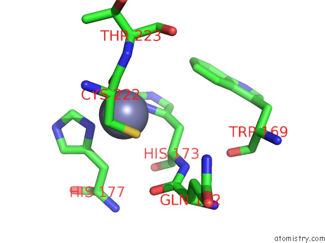
Mono view
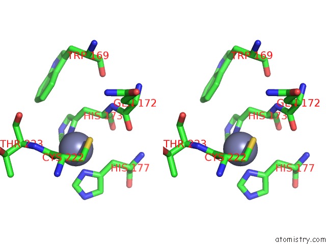
Stereo pair view
Mono view
Stereo pair view
A full contact list of Zinc with other atoms in the Zn binding
site number 1 of The Value 'Crystal Structure of Fad Quinone Reductase 2 at 1.45A within 5.0Å range:
|
Zinc binding site 2 out of 2 in 4qoe
Go back to
Zinc binding site 2 out
of 2 in the The Value 'Crystal Structure of Fad Quinone Reductase 2 at 1.45A
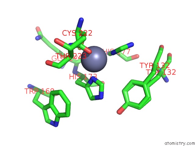
Mono view
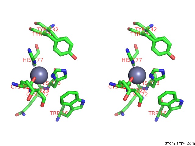
Stereo pair view
Mono view
Stereo pair view
A full contact list of Zinc with other atoms in the Zn binding
site number 2 of The Value 'Crystal Structure of Fad Quinone Reductase 2 at 1.45A within 5.0Å range:
|
Reference:
J.Serriere,
J.A.Boutin,
T.Isabet,
M.Antoine,
G.Ferry.
The Value 'Crystal Structure of Fad Quinone Reductase 2 at 1.45A To Be Published.
Page generated: Wed Aug 20 21:52:05 2025
Last articles
Zn in 5YI3Zn in 5YIQ
Zn in 5YIX
Zn in 5YI0
Zn in 5YHZ
Zn in 5YHY
Zn in 5YHX
Zn in 5YHT
Zn in 5YEL
Zn in 5YEF