Zinc »
PDB 4mtp-4n4e »
4mxk »
Zinc in PDB 4mxk: X-Ray Structure of Fe(II)-ZNPIXFEBMB1
Protein crystallography data
The structure of X-Ray Structure of Fe(II)-ZNPIXFEBMB1, PDB code: 4mxk
was solved by
S.Chakraborty,
Y.Lu,
I.Petrik,
with X-Ray Crystallography technique. A brief refinement statistics is given in the table below:
| Resolution Low / High (Å) | 40.34 / 1.52 |
| Space group | P 21 21 21 |
| Cell size a, b, c (Å), α, β, γ (°) | 39.750, 47.593, 76.008, 90.00, 90.00, 90.00 |
| R / Rfree (%) | 17.9 / 22 |
Other elements in 4mxk:
The structure of X-Ray Structure of Fe(II)-ZNPIXFEBMB1 also contains other interesting chemical elements:
| Iron | (Fe) | 2 atoms |
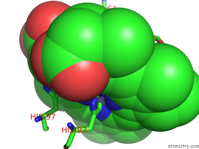
Zinc Binding Sites:
The binding sites of Zinc atom in the X-Ray Structure of Fe(II)-ZNPIXFEBMB1
(pdb code 4mxk). This binding sites where shown within
5.0 Angstroms radius around Zinc atom.
In total only one binding site of Zinc was determined in the X-Ray Structure of Fe(II)-ZNPIXFEBMB1, PDB code: 4mxk:
In total only one binding site of Zinc was determined in the X-Ray Structure of Fe(II)-ZNPIXFEBMB1, PDB code: 4mxk:
Zinc binding site 1 out of 1 in 4mxk
Go back to
Zinc binding site 1 out
of 1 in the X-Ray Structure of Fe(II)-ZNPIXFEBMB1
Mono view
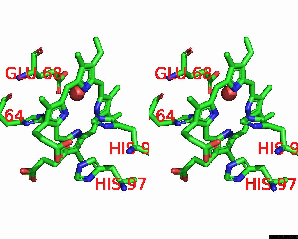
Stereo pair view
Mono view
Stereo pair view
A full contact list of Zinc with other atoms in the Zn binding
site number 1 of X-Ray Structure of Fe(II)-ZNPIXFEBMB1 within 5.0Å range:
|
Reference:
S.Chakraborty,
J.Reed,
M.Ross,
M.J.Nilges,
I.D.Petrik,
S.Ghosh,
S.Hammes-Schiffer,
J.T.Sage,
Y.Zhang,
C.E.Schulz,
Y.Lu.
Spectroscopic and Computational Study of A Nonheme Iron Nitrosyl Center in A Biosynthetic Model of Nitric Oxide Reductase. Angew.Chem.Int.Ed.Engl. V. 53 2417 2014.
ISSN: ISSN 1433-7851
PubMed: 24481708
DOI: 10.1002/ANIE.201308431
Page generated: Wed Aug 20 20:35:35 2025
ISSN: ISSN 1433-7851
PubMed: 24481708
DOI: 10.1002/ANIE.201308431
Last articles
Zn in 5CEHZn in 5CE5
Zn in 5CCV
Zn in 5CDT
Zn in 5CDS
Zn in 5CDE
Zn in 5CDG
Zn in 5CBM
Zn in 5CD4
Zn in 5CC1