Zinc »
PDB 4eg2-4eyp »
4ej6 »
Zinc in PDB 4ej6: Crystal Structure of A Putative Zinc-Binding Dehydrogenase (Target Psi-012003) From Sinorhizobium Meliloti 1021
Protein crystallography data
The structure of Crystal Structure of A Putative Zinc-Binding Dehydrogenase (Target Psi-012003) From Sinorhizobium Meliloti 1021, PDB code: 4ej6
was solved by
P.Sampathkumar,
S.C.Almo,
New York Structural Genomics Researchconsortium (Nysgrc),
with X-Ray Crystallography technique. A brief refinement statistics is given in the table below:
| Resolution Low / High (Å) | 50.00 / 1.89 |
| Space group | I 4 2 2 |
| Cell size a, b, c (Å), α, β, γ (°) | 107.607, 107.607, 137.160, 90.00, 90.00, 90.00 |
| R / Rfree (%) | 17.6 / 20.2 |
Other elements in 4ej6:
The structure of Crystal Structure of A Putative Zinc-Binding Dehydrogenase (Target Psi-012003) From Sinorhizobium Meliloti 1021 also contains other interesting chemical elements:
| Chlorine | (Cl) | 1 atom |
Zinc Binding Sites:
The binding sites of Zinc atom in the Crystal Structure of A Putative Zinc-Binding Dehydrogenase (Target Psi-012003) From Sinorhizobium Meliloti 1021
(pdb code 4ej6). This binding sites where shown within
5.0 Angstroms radius around Zinc atom.
In total only one binding site of Zinc was determined in the Crystal Structure of A Putative Zinc-Binding Dehydrogenase (Target Psi-012003) From Sinorhizobium Meliloti 1021, PDB code: 4ej6:
In total only one binding site of Zinc was determined in the Crystal Structure of A Putative Zinc-Binding Dehydrogenase (Target Psi-012003) From Sinorhizobium Meliloti 1021, PDB code: 4ej6:
Zinc binding site 1 out of 1 in 4ej6
Go back to
Zinc binding site 1 out
of 1 in the Crystal Structure of A Putative Zinc-Binding Dehydrogenase (Target Psi-012003) From Sinorhizobium Meliloti 1021
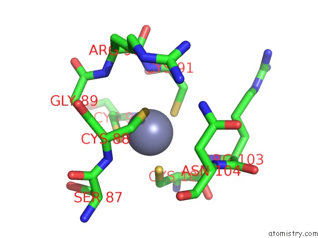
Mono view
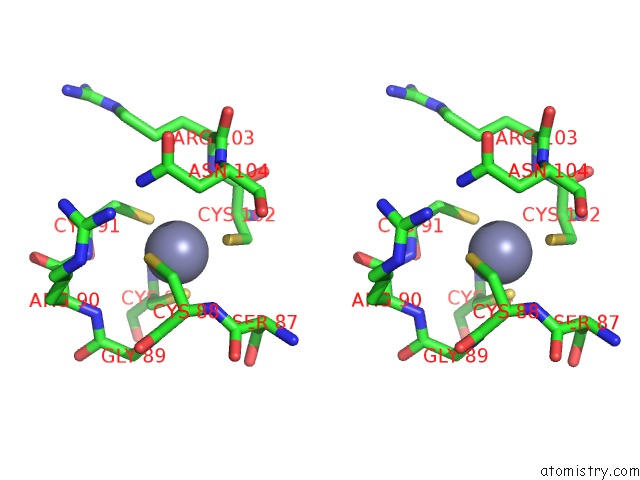
Stereo pair view
Mono view
Stereo pair view
A full contact list of Zinc with other atoms in the Zn binding
site number 1 of Crystal Structure of A Putative Zinc-Binding Dehydrogenase (Target Psi-012003) From Sinorhizobium Meliloti 1021 within 5.0Å range:
|
Reference:
P.Sampathkumar,
N.Banu,
R.Bhosle,
J.Bonanno,
S.Chamala,
S.Chowdhury,
A.Fiser,
A.Gizzi,
A.S.Glenn,
J.Hammonds,
B.Hillerich,
K.Khafizov,
J.D.Love,
B.Matikainen,
Y.Patskovsky,
R.Seidel,
R.Toro,
W.Zencheck,
S.C.Almo.
Crystal Structure of A Putative Zinc-Binding Dehydrogenase From Sinorhizobium Meliloti 1021 To Be Published.
Page generated: Wed Aug 20 17:25:23 2025
Last articles
Zn in 6ACLZn in 6ACE
Zn in 6AA5
Zn in 6ACB
Zn in 6AA3
Zn in 6AA4
Zn in 6A8Z
Zn in 6A94
Zn in 6A8L
Zn in 6A93