Zinc »
PDB 4eg2-4eyp »
4ej5 »
Zinc in PDB 4ej5: Crystal Structure of the Catalytic Domain of Botulinum Neurotoxin Bont/A Wild-Type
Enzymatic activity of Crystal Structure of the Catalytic Domain of Botulinum Neurotoxin Bont/A Wild-Type
All present enzymatic activity of Crystal Structure of the Catalytic Domain of Botulinum Neurotoxin Bont/A Wild-Type:
3.4.24.69;
3.4.24.69;
Protein crystallography data
The structure of Crystal Structure of the Catalytic Domain of Botulinum Neurotoxin Bont/A Wild-Type, PDB code: 4ej5
was solved by
E.A.Stura,
L.Vera,
V.Dive,
with X-Ray Crystallography technique. A brief refinement statistics is given in the table below:
| Resolution Low / High (Å) | 42.17 / 1.87 |
| Space group | P 1 21 1 |
| Cell size a, b, c (Å), α, β, γ (°) | 49.950, 66.370, 64.720, 90.00, 98.24, 90.00 |
| R / Rfree (%) | 16.1 / 20.8 |
Zinc Binding Sites:
The binding sites of Zinc atom in the Crystal Structure of the Catalytic Domain of Botulinum Neurotoxin Bont/A Wild-Type
(pdb code 4ej5). This binding sites where shown within
5.0 Angstroms radius around Zinc atom.
In total only one binding site of Zinc was determined in the Crystal Structure of the Catalytic Domain of Botulinum Neurotoxin Bont/A Wild-Type, PDB code: 4ej5:
In total only one binding site of Zinc was determined in the Crystal Structure of the Catalytic Domain of Botulinum Neurotoxin Bont/A Wild-Type, PDB code: 4ej5:
Zinc binding site 1 out of 1 in 4ej5
Go back to
Zinc binding site 1 out
of 1 in the Crystal Structure of the Catalytic Domain of Botulinum Neurotoxin Bont/A Wild-Type
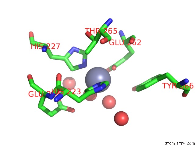
Mono view
Stereo pair view
Mono view
Stereo pair view
A full contact list of Zinc with other atoms in the Zn binding
site number 1 of Crystal Structure of the Catalytic Domain of Botulinum Neurotoxin Bont/A Wild-Type within 5.0Å range:
|
Reference:
E.A.Stura,
L.Le Roux,
K.Guitot,
S.Garcia,
S.Bregant,
F.Beau,
L.Vera,
G.Collet,
D.Ptchelkine,
H.Bakirci,
V.Dive.
Structural Framework For Covalent Inhibition of Clostridium Botulinum Neurotoxin A By Targeting CYS165. J.Biol.Chem. V. 287 33607 2012.
ISSN: ISSN 0021-9258
PubMed: 22869371
DOI: 10.1074/JBC.M112.396697
Page generated: Wed Aug 20 17:25:13 2025
ISSN: ISSN 0021-9258
PubMed: 22869371
DOI: 10.1074/JBC.M112.396697
Last articles
Zn in 6A5MZn in 6A5L
Zn in 6A5N
Zn in 6A5K
Zn in 6A59
Zn in 6A58
Zn in 6A57
Zn in 5ZYB
Zn in 5ZZU
Zn in 6A3Z