Zinc »
PDB 3krv-3l2u »
3l1u »
Zinc in PDB 3l1u: Crystal Structure of Calcium-Bound Gmhb From E. Coli.
Protein crystallography data
The structure of Crystal Structure of Calcium-Bound Gmhb From E. Coli., PDB code: 3l1u
was solved by
S.N.Sugiman-Marangos,
M.S.Junop,
with X-Ray Crystallography technique. A brief refinement statistics is given in the table below:
| Resolution Low / High (Å) | 45.57 / 1.95 |
| Space group | P 21 21 21 |
| Cell size a, b, c (Å), α, β, γ (°) | 50.553, 64.031, 105.236, 90.00, 90.00, 90.00 |
| R / Rfree (%) | 23.5 / 25.8 |
Other elements in 3l1u:
The structure of Crystal Structure of Calcium-Bound Gmhb From E. Coli. also contains other interesting chemical elements:
| Calcium | (Ca) | 2 atoms |
Zinc Binding Sites:
The binding sites of Zinc atom in the Crystal Structure of Calcium-Bound Gmhb From E. Coli.
(pdb code 3l1u). This binding sites where shown within
5.0 Angstroms radius around Zinc atom.
In total 2 binding sites of Zinc where determined in the Crystal Structure of Calcium-Bound Gmhb From E. Coli., PDB code: 3l1u:
Jump to Zinc binding site number: 1; 2;
In total 2 binding sites of Zinc where determined in the Crystal Structure of Calcium-Bound Gmhb From E. Coli., PDB code: 3l1u:
Jump to Zinc binding site number: 1; 2;
Zinc binding site 1 out of 2 in 3l1u
Go back to
Zinc binding site 1 out
of 2 in the Crystal Structure of Calcium-Bound Gmhb From E. Coli.
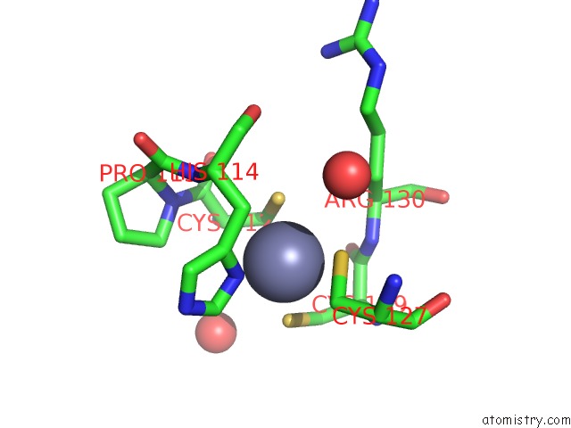
Mono view
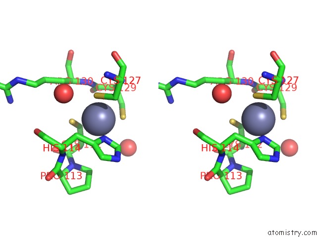
Stereo pair view
Mono view
Stereo pair view
A full contact list of Zinc with other atoms in the Zn binding
site number 1 of Crystal Structure of Calcium-Bound Gmhb From E. Coli. within 5.0Å range:
|
Zinc binding site 2 out of 2 in 3l1u
Go back to
Zinc binding site 2 out
of 2 in the Crystal Structure of Calcium-Bound Gmhb From E. Coli.
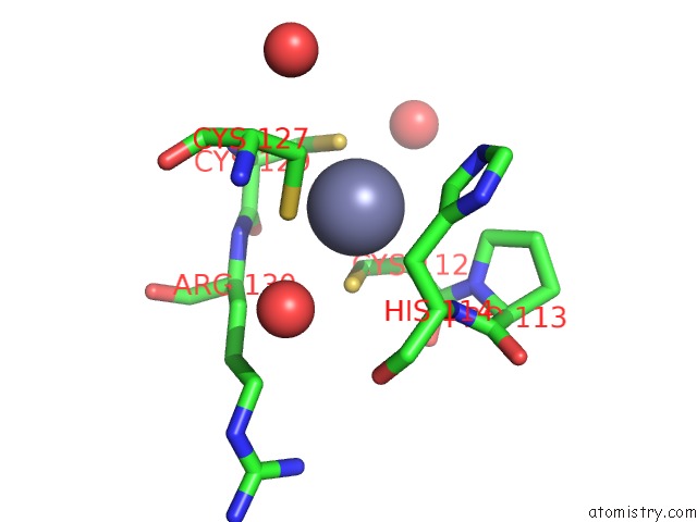
Mono view
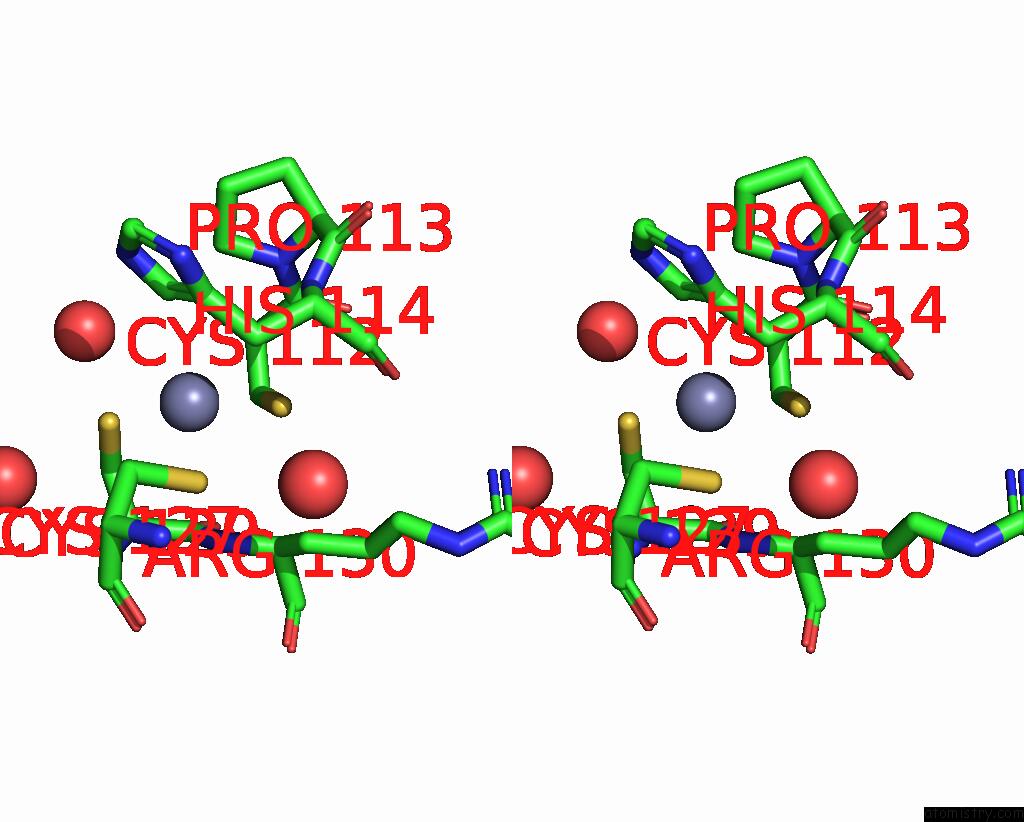
Stereo pair view
Mono view
Stereo pair view
A full contact list of Zinc with other atoms in the Zn binding
site number 2 of Crystal Structure of Calcium-Bound Gmhb From E. Coli. within 5.0Å range:
|
Reference:
P.L.Taylor,
S.Sugiman-Marangos,
K.Zhang,
M.A.Valvano,
G.D.Wright,
M.S.Junop.
Structural and Kinetic Characterization of the Lps Biosynthetic Enzyme D-Alpha,Beta-D-Heptose-1,7-Bisphosphate Phosphatase (Gmhb) From Escherichia Coli. Biochemistry V. 49 1033 2010.
ISSN: ISSN 0006-2960
PubMed: 20050699
DOI: 10.1021/BI901780J
Page generated: Sat Oct 26 08:21:03 2024
ISSN: ISSN 0006-2960
PubMed: 20050699
DOI: 10.1021/BI901780J
Last articles
Fe in 2YXOFe in 2YRS
Fe in 2YXC
Fe in 2YNM
Fe in 2YVJ
Fe in 2YP1
Fe in 2YU2
Fe in 2YU1
Fe in 2YQB
Fe in 2YOO