Zinc »
PDB 3gtv-3h68 »
3h5r »
Zinc in PDB 3h5r: Crystal Structure of E. Coli Mccb + Succinimide
Protein crystallography data
The structure of Crystal Structure of E. Coli Mccb + Succinimide, PDB code: 3h5r
was solved by
C.A.Regni,
R.F.Roush,
D.Miller,
A.Nourse,
C.T.Walsh,
B.A.Schulman,
with X-Ray Crystallography technique. A brief refinement statistics is given in the table below:
| Resolution Low / High (Å) | 27.95 / 2.10 |
| Space group | P 1 21 1 |
| Cell size a, b, c (Å), α, β, γ (°) | 55.934, 137.972, 80.134, 90.00, 92.10, 90.00 |
| R / Rfree (%) | 19.4 / 25 |
Zinc Binding Sites:
The binding sites of Zinc atom in the Crystal Structure of E. Coli Mccb + Succinimide
(pdb code 3h5r). This binding sites where shown within
5.0 Angstroms radius around Zinc atom.
In total 4 binding sites of Zinc where determined in the Crystal Structure of E. Coli Mccb + Succinimide, PDB code: 3h5r:
Jump to Zinc binding site number: 1; 2; 3; 4;
In total 4 binding sites of Zinc where determined in the Crystal Structure of E. Coli Mccb + Succinimide, PDB code: 3h5r:
Jump to Zinc binding site number: 1; 2; 3; 4;
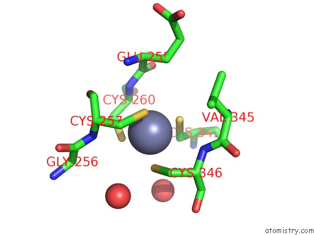
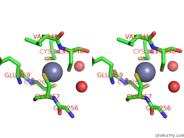
Zinc binding site 1 out of 4 in 3h5r
Go back to
Zinc binding site 1 out
of 4 in the Crystal Structure of E. Coli Mccb + Succinimide
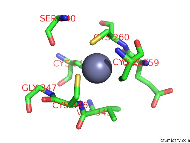
Mono view
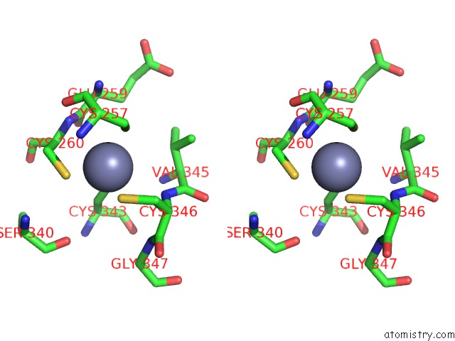
Stereo pair view
Mono view
Stereo pair view
A full contact list of Zinc with other atoms in the Zn binding
site number 1 of Crystal Structure of E. Coli Mccb + Succinimide within 5.0Å range:
|
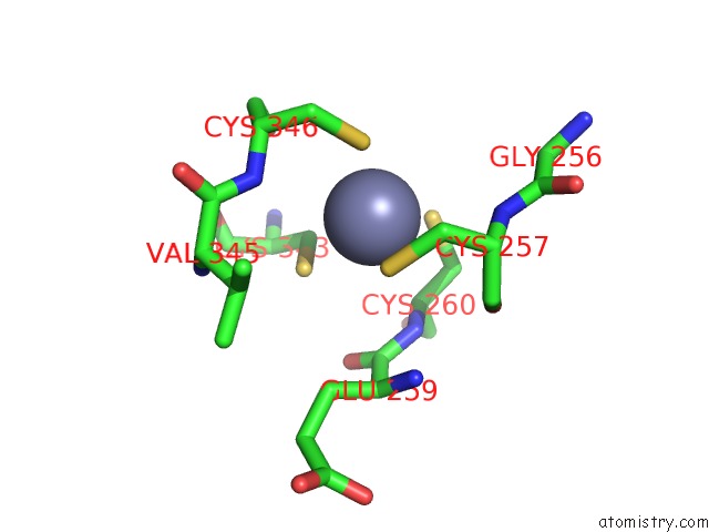
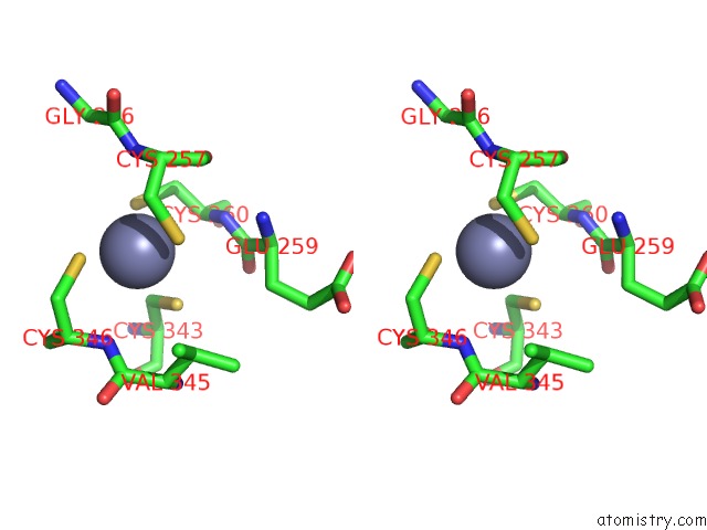
Zinc binding site 2 out of 4 in 3h5r
Go back to
Zinc binding site 2 out
of 4 in the Crystal Structure of E. Coli Mccb + Succinimide
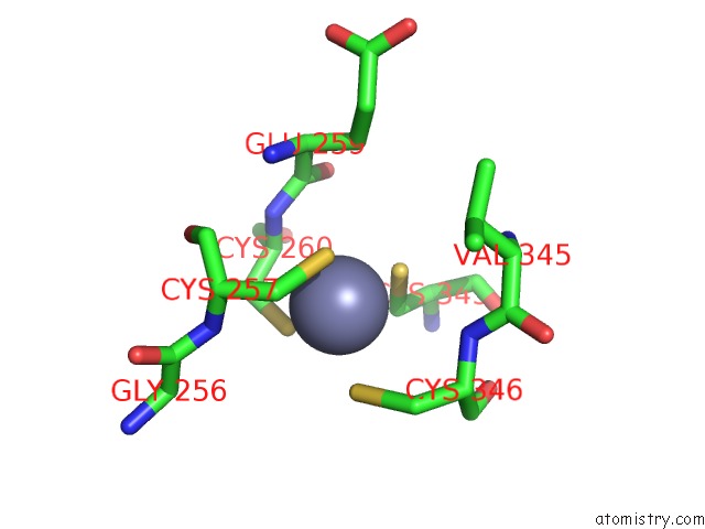
Mono view
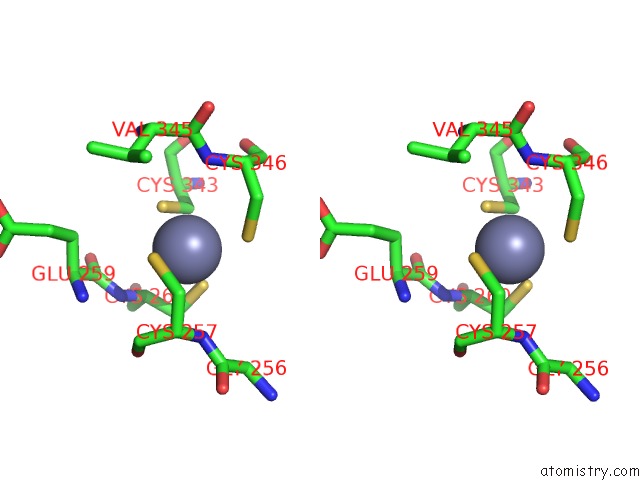
Stereo pair view
Mono view
Stereo pair view
A full contact list of Zinc with other atoms in the Zn binding
site number 2 of Crystal Structure of E. Coli Mccb + Succinimide within 5.0Å range:
|
Zinc binding site 3 out of 4 in 3h5r
Go back to
Zinc binding site 3 out
of 4 in the Crystal Structure of E. Coli Mccb + Succinimide
Mono view
Stereo pair view
Mono view
Stereo pair view
A full contact list of Zinc with other atoms in the Zn binding
site number 3 of Crystal Structure of E. Coli Mccb + Succinimide within 5.0Å range:
|
Zinc binding site 4 out of 4 in 3h5r
Go back to
Zinc binding site 4 out
of 4 in the Crystal Structure of E. Coli Mccb + Succinimide
Mono view
Stereo pair view
Mono view
Stereo pair view
A full contact list of Zinc with other atoms in the Zn binding
site number 4 of Crystal Structure of E. Coli Mccb + Succinimide within 5.0Å range:
|
Reference:
C.A.Regni,
R.F.Roush,
D.J.Miller,
A.Nourse,
C.T.Walsh,
B.A.Schulman.
How the Mccb Bacterial Ancestor of Ubiquitin E1 Initiates Biosynthesis of the Microcin C7 Antibiotic. Embo J. V. 28 1953 2009.
ISSN: ISSN 0261-4189
PubMed: 19494832
DOI: 10.1038/EMBOJ.2009.146
Page generated: Thu Oct 24 14:14:42 2024
ISSN: ISSN 0261-4189
PubMed: 19494832
DOI: 10.1038/EMBOJ.2009.146
Last articles
Mg in 5OEAMg in 5OET
Mg in 5OED
Mg in 5OEB
Mg in 5OEC
Mg in 5ODZ
Mg in 5ODO
Mg in 5ODC
Mg in 5ODJ
Mg in 5OCT