Zinc »
PDB 3gtv-3h68 »
3h15 »
Zinc in PDB 3h15: Crystal Structure of Replication Initiation Factor MCM10-Id Bound to Ssdna
Protein crystallography data
The structure of Crystal Structure of Replication Initiation Factor MCM10-Id Bound to Ssdna, PDB code: 3h15
was solved by
E.M.Warren,
B.F.Eichman,
with X-Ray Crystallography technique. A brief refinement statistics is given in the table below:
| Resolution Low / High (Å) | 34.14 / 2.72 |
| Space group | P 31 2 1 |
| Cell size a, b, c (Å), α, β, γ (°) | 95.017, 95.017, 61.159, 90.00, 90.00, 120.00 |
| R / Rfree (%) | 19.7 / 23.2 |
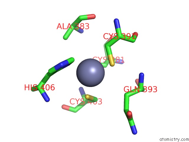
Zinc Binding Sites:
The binding sites of Zinc atom in the Crystal Structure of Replication Initiation Factor MCM10-Id Bound to Ssdna
(pdb code 3h15). This binding sites where shown within
5.0 Angstroms radius around Zinc atom.
In total only one binding site of Zinc was determined in the Crystal Structure of Replication Initiation Factor MCM10-Id Bound to Ssdna, PDB code: 3h15:
In total only one binding site of Zinc was determined in the Crystal Structure of Replication Initiation Factor MCM10-Id Bound to Ssdna, PDB code: 3h15:
Zinc binding site 1 out of 1 in 3h15
Go back to
Zinc binding site 1 out
of 1 in the Crystal Structure of Replication Initiation Factor MCM10-Id Bound to Ssdna
Mono view
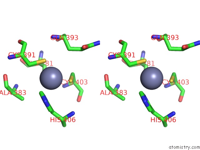
Stereo pair view
Mono view
Stereo pair view
A full contact list of Zinc with other atoms in the Zn binding
site number 1 of Crystal Structure of Replication Initiation Factor MCM10-Id Bound to Ssdna within 5.0Å range:
|
Reference:
E.M.Warren,
H.Huang,
E.Fanning,
W.J.Chazin,
B.F.Eichman.
Physical Interactions Between MCM10, Dna, and Dna Polymerase {Alpha}. J.Biol.Chem. V. 284 24662 2009.
ISSN: ISSN 0021-9258
PubMed: 19608746
DOI: 10.1074/JBC.M109.020438
Page generated: Thu Oct 24 14:10:30 2024
ISSN: ISSN 0021-9258
PubMed: 19608746
DOI: 10.1074/JBC.M109.020438
Last articles
Mg in 6BBZMg in 6BBR
Mg in 6BBQ
Mg in 6BBP
Mg in 6BA7
Mg in 6BBL
Mg in 6B9G
Mg in 6B8H
Mg in 6B9F
Mg in 6B9E