Zinc »
PDB 3dsx-3e2i »
3e1z »
Zinc in PDB 3e1z: Crystal Structure of the Parasite Protesase Inhibitor Chagasin in Complex with Papain
Enzymatic activity of Crystal Structure of the Parasite Protesase Inhibitor Chagasin in Complex with Papain
All present enzymatic activity of Crystal Structure of the Parasite Protesase Inhibitor Chagasin in Complex with Papain:
3.4.22.2;
3.4.22.2;
Protein crystallography data
The structure of Crystal Structure of the Parasite Protesase Inhibitor Chagasin in Complex with Papain, PDB code: 3e1z
was solved by
I.Redzynia,
G.Bujacz,
A.Bujacz,
A.Ljunggren,
M.Abrahamson,
M.Jaskolski,
with X-Ray Crystallography technique. A brief refinement statistics is given in the table below:
| Resolution Low / High (Å) | 60.00 / 1.86 |
| Space group | I 4 2 2 |
| Cell size a, b, c (Å), α, β, γ (°) | 99.130, 99.130, 159.490, 90.00, 90.00, 90.00 |
| R / Rfree (%) | 16.4 / 20.8 |
Zinc Binding Sites:
The binding sites of Zinc atom in the Crystal Structure of the Parasite Protesase Inhibitor Chagasin in Complex with Papain
(pdb code 3e1z). This binding sites where shown within
5.0 Angstroms radius around Zinc atom.
In total only one binding site of Zinc was determined in the Crystal Structure of the Parasite Protesase Inhibitor Chagasin in Complex with Papain, PDB code: 3e1z:
In total only one binding site of Zinc was determined in the Crystal Structure of the Parasite Protesase Inhibitor Chagasin in Complex with Papain, PDB code: 3e1z:
Zinc binding site 1 out of 1 in 3e1z
Go back to
Zinc binding site 1 out
of 1 in the Crystal Structure of the Parasite Protesase Inhibitor Chagasin in Complex with Papain
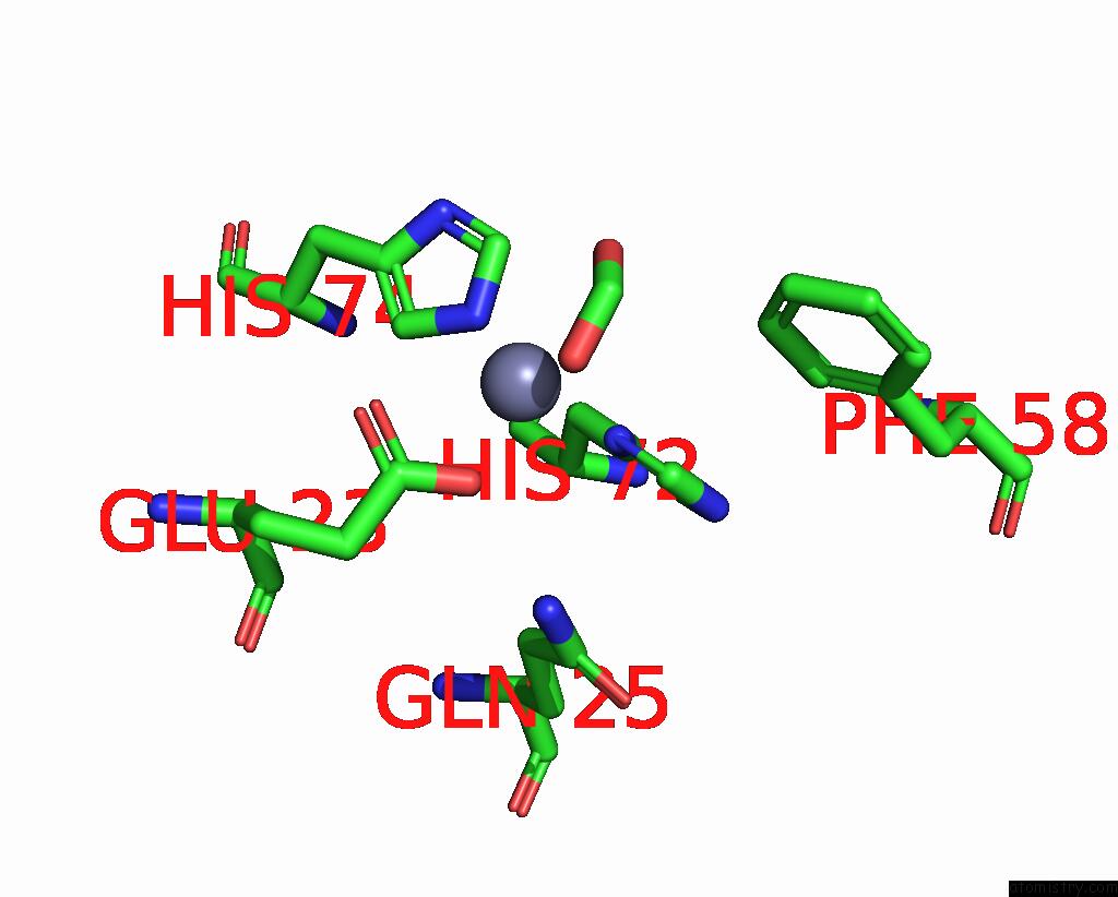
Mono view
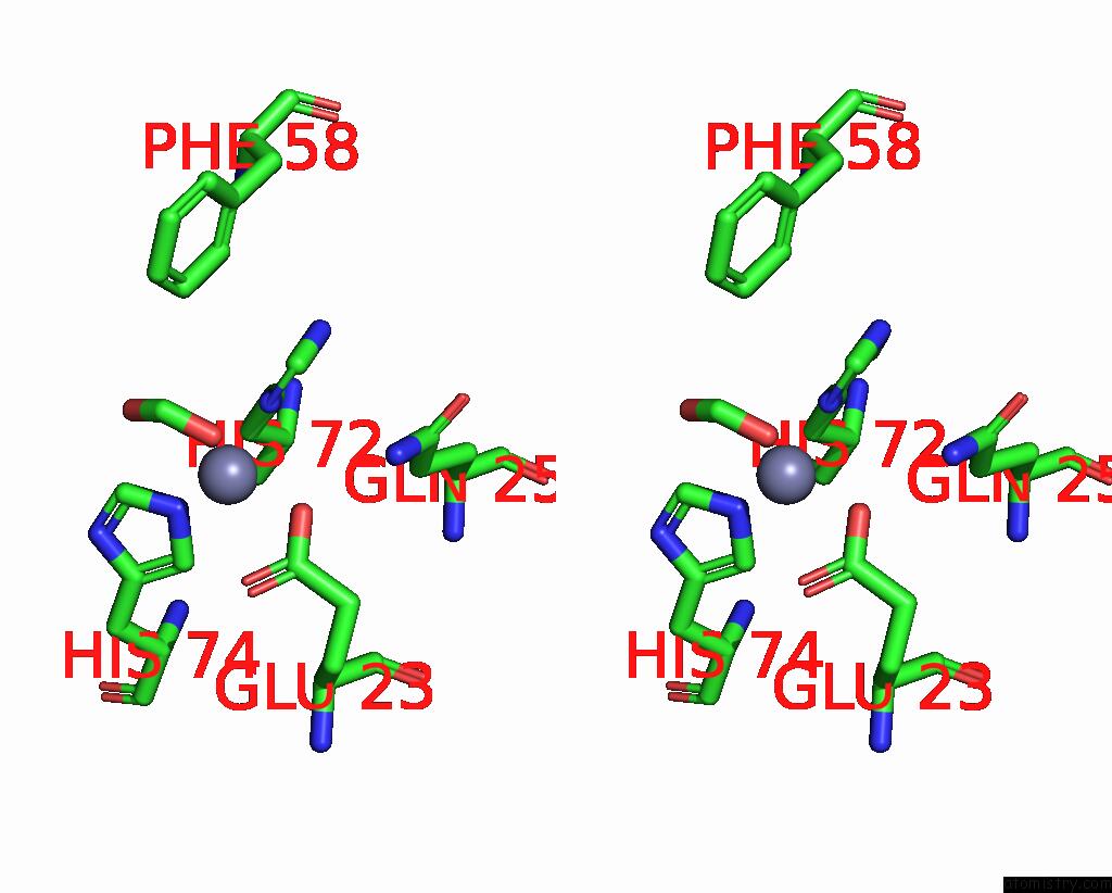
Stereo pair view
Mono view
Stereo pair view
A full contact list of Zinc with other atoms in the Zn binding
site number 1 of Crystal Structure of the Parasite Protesase Inhibitor Chagasin in Complex with Papain within 5.0Å range:
|
Reference:
I.Redzynia,
A.Ljunggren,
A.Bujacz,
M.Abrahamson,
M.Jaskolski,
G.Bujacz.
Crystal Structure of the Parasite Inhibitor Chagasin in Complex with Papain Allows Identification of Structural Requirements For Broad Reactivity and Specificity Determinants For Target Proteases. Febs J. V. 276 793 2009.
ISSN: ISSN 1742-464X
PubMed: 19143838
DOI: 10.1111/J.1742-4658.2008.06824.X
Page generated: Thu Oct 24 12:28:52 2024
ISSN: ISSN 1742-464X
PubMed: 19143838
DOI: 10.1111/J.1742-4658.2008.06824.X
Last articles
Mg in 5QJIMg in 5QJJ
Mg in 5QJH
Mg in 5QJG
Mg in 5QJD
Mg in 5QJF
Mg in 5QJE
Mg in 5QJC
Mg in 5QJB
Mg in 5QJ9