Zinc »
PDB 3d7v-3dgm »
3dbk »
Zinc in PDB 3dbk: Pseudomonas Aeruginosa Elastase with Phosphoramidon
Protein crystallography data
The structure of Pseudomonas Aeruginosa Elastase with Phosphoramidon, PDB code: 3dbk
was solved by
D.B.Mckay,
M.T.Overgaard,
with X-Ray Crystallography technique. A brief refinement statistics is given in the table below:
| Resolution Low / High (Å) | 29.17 / 1.40 |
| Space group | P 21 21 21 |
| Cell size a, b, c (Å), α, β, γ (°) | 44.000, 50.750, 121.510, 90.00, 90.00, 90.00 |
| R / Rfree (%) | 18.3 / 20.3 |
Other elements in 3dbk:
The structure of Pseudomonas Aeruginosa Elastase with Phosphoramidon also contains other interesting chemical elements:
| Calcium | (Ca) | 1 atom |
Zinc Binding Sites:
The binding sites of Zinc atom in the Pseudomonas Aeruginosa Elastase with Phosphoramidon
(pdb code 3dbk). This binding sites where shown within
5.0 Angstroms radius around Zinc atom.
In total only one binding site of Zinc was determined in the Pseudomonas Aeruginosa Elastase with Phosphoramidon, PDB code: 3dbk:
In total only one binding site of Zinc was determined in the Pseudomonas Aeruginosa Elastase with Phosphoramidon, PDB code: 3dbk:
Zinc binding site 1 out of 1 in 3dbk
Go back to
Zinc binding site 1 out
of 1 in the Pseudomonas Aeruginosa Elastase with Phosphoramidon
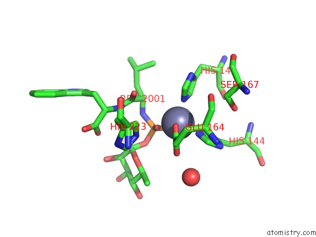
Mono view
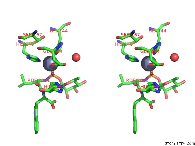
Stereo pair view
Mono view
Stereo pair view
A full contact list of Zinc with other atoms in the Zn binding
site number 1 of Pseudomonas Aeruginosa Elastase with Phosphoramidon within 5.0Å range:
|
Reference:
M.T.Overgaard,
D.B.Mckay.
Structure of the Elastase of Pseudomonas Aeruginosa Complexed with Phosphoramidon To Be Published.
Page generated: Thu Oct 24 12:07:06 2024
Last articles
Mg in 4LF7Mg in 4LF6
Mg in 4LF4
Mg in 4LF5
Mg in 4LCZ
Mg in 4LF2
Mg in 4LF1
Mg in 4LEM
Mg in 4LCK
Mg in 4LE0