Zinc »
PDB 3c52-3cjj »
3caj »
Zinc in PDB 3caj: Crystal Structure of the Human Carbonic Anhydrase II in Complex with Ethoxzolamide
Enzymatic activity of Crystal Structure of the Human Carbonic Anhydrase II in Complex with Ethoxzolamide
All present enzymatic activity of Crystal Structure of the Human Carbonic Anhydrase II in Complex with Ethoxzolamide:
4.2.1.1;
4.2.1.1;
Protein crystallography data
The structure of Crystal Structure of the Human Carbonic Anhydrase II in Complex with Ethoxzolamide, PDB code: 3caj
was solved by
A.Di Fiore,
G.De Simone,
with X-Ray Crystallography technique. A brief refinement statistics is given in the table below:
| Resolution Low / High (Å) | 20.00 / 1.80 |
| Space group | P 1 21 1 |
| Cell size a, b, c (Å), α, β, γ (°) | 42.210, 41.360, 72.030, 90.00, 104.35, 90.00 |
| R / Rfree (%) | 18 / 21.4 |
Other elements in 3caj:
The structure of Crystal Structure of the Human Carbonic Anhydrase II in Complex with Ethoxzolamide also contains other interesting chemical elements:
| Mercury | (Hg) | 1 atom |
| Chlorine | (Cl) | 1 atom |
Zinc Binding Sites:
The binding sites of Zinc atom in the Crystal Structure of the Human Carbonic Anhydrase II in Complex with Ethoxzolamide
(pdb code 3caj). This binding sites where shown within
5.0 Angstroms radius around Zinc atom.
In total only one binding site of Zinc was determined in the Crystal Structure of the Human Carbonic Anhydrase II in Complex with Ethoxzolamide, PDB code: 3caj:
In total only one binding site of Zinc was determined in the Crystal Structure of the Human Carbonic Anhydrase II in Complex with Ethoxzolamide, PDB code: 3caj:
Zinc binding site 1 out of 1 in 3caj
Go back to
Zinc binding site 1 out
of 1 in the Crystal Structure of the Human Carbonic Anhydrase II in Complex with Ethoxzolamide
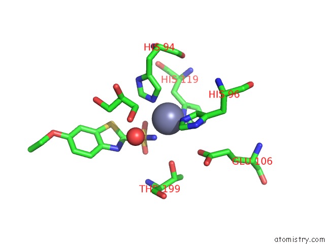
Mono view
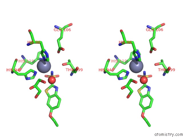
Stereo pair view
Mono view
Stereo pair view
A full contact list of Zinc with other atoms in the Zn binding
site number 1 of Crystal Structure of the Human Carbonic Anhydrase II in Complex with Ethoxzolamide within 5.0Å range:
|
Reference:
A.Di Fiore,
C.Pedone,
J.Antel,
H.Waldeck,
A.Witte,
M.Wurl,
A.Scozzafava,
C.T.Supuran,
G.De Simone.
Carbonic Anhydrase Inhibitors: the X-Ray Crystal Structure of Ethoxzolamide Complexed to Human Isoform II Reveals the Importance of THR200 and GLN92 For Obtaining Tight-Binding Inhibitors Bioorg.Med.Chem.Lett. V. 18 2669 2008.
ISSN: ISSN 0960-894X
PubMed: 18359629
DOI: 10.1016/J.BMCL.2008.03.023
Page generated: Thu Oct 24 11:45:55 2024
ISSN: ISSN 0960-894X
PubMed: 18359629
DOI: 10.1016/J.BMCL.2008.03.023
Last articles
Mg in 9AZLMg in 9AZ6
Mg in 9AZ5
Mg in 9AZ4
Mg in 9AYO
Mg in 9AYP
Mg in 9AW3
Mg in 9AYN
Mg in 9AYI
Mg in 9AXL