Zinc »
PDB 2y7g-2yqp »
2yb5 »
Zinc in PDB 2yb5: Structure of the Fusidic Acid Resistance Protein Fusc
Protein crystallography data
The structure of Structure of the Fusidic Acid Resistance Protein Fusc, PDB code: 2yb5
was solved by
G.Cox,
T.A.Edwards,
with X-Ray Crystallography technique. A brief refinement statistics is given in the table below:
| Resolution Low / High (Å) | 15.00 / 2.10 |
| Space group | P 1 21 1 |
| Cell size a, b, c (Å), α, β, γ (°) | 34.150, 109.610, 61.150, 90.00, 101.72, 90.00 |
| R / Rfree (%) | 21.334 / 28.773 |
Zinc Binding Sites:
The binding sites of Zinc atom in the Structure of the Fusidic Acid Resistance Protein Fusc
(pdb code 2yb5). This binding sites where shown within
5.0 Angstroms radius around Zinc atom.
In total 2 binding sites of Zinc where determined in the Structure of the Fusidic Acid Resistance Protein Fusc, PDB code: 2yb5:
Jump to Zinc binding site number: 1; 2;
In total 2 binding sites of Zinc where determined in the Structure of the Fusidic Acid Resistance Protein Fusc, PDB code: 2yb5:
Jump to Zinc binding site number: 1; 2;
Zinc binding site 1 out of 2 in 2yb5
Go back to
Zinc binding site 1 out
of 2 in the Structure of the Fusidic Acid Resistance Protein Fusc
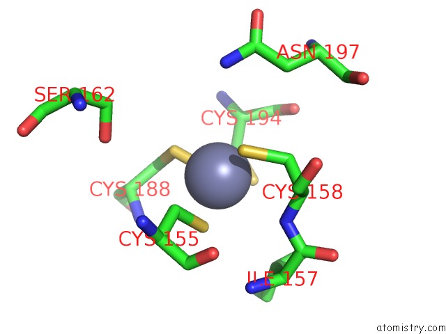
Mono view
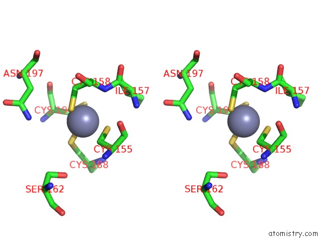
Stereo pair view
Mono view
Stereo pair view
A full contact list of Zinc with other atoms in the Zn binding
site number 1 of Structure of the Fusidic Acid Resistance Protein Fusc within 5.0Å range:
|
Zinc binding site 2 out of 2 in 2yb5
Go back to
Zinc binding site 2 out
of 2 in the Structure of the Fusidic Acid Resistance Protein Fusc

Mono view

Stereo pair view
Mono view
Stereo pair view
A full contact list of Zinc with other atoms in the Zn binding
site number 2 of Structure of the Fusidic Acid Resistance Protein Fusc within 5.0Å range:
|
Reference:
G.Cox,
G.S.Thompson,
H.T.Jenkins,
S.W.Homans,
T.A.Edwards,
A.J.Oneill.
Ribosome Clearance By Fusb-Type Proteins Mediates Resistance to the Antibiotic Fusidic Acid Proc.Natl.Acad.Sci.Usa V. 109 2102 2012.
ISSN: ISSN 0027-8424
PubMed: 22308410
DOI: 10.1073/PNAS.1117275109
Page generated: Thu Oct 17 05:47:36 2024
ISSN: ISSN 0027-8424
PubMed: 22308410
DOI: 10.1073/PNAS.1117275109
Last articles
F in 5W4VF in 5W8I
F in 5W8B
F in 5W77
F in 5W4W
F in 5W5W
F in 5W58
F in 5W2Y
F in 5W2W
F in 5W2U