Zinc »
PDB 8woq-8x2i »
8wtc »
Zinc in PDB 8wtc: Crystal Structure of Mcsb Kinase Domain Complexed with Mcsa.
Protein crystallography data
The structure of Crystal Structure of Mcsb Kinase Domain Complexed with Mcsa., PDB code: 8wtc
was solved by
M.Arifuzzaman,
E.Kwon,
D.Y.Kim,
with X-Ray Crystallography technique. A brief refinement statistics is given in the table below:
| Resolution Low / High (Å) | 43.45 / 2.80 |
| Space group | C 1 2 1 |
| Cell size a, b, c (Å), α, β, γ (°) | 188.104, 74.208, 79.935, 90, 100.84, 90 |
| R / Rfree (%) | 21.6 / 26.6 |
Zinc Binding Sites:
The binding sites of Zinc atom in the Crystal Structure of Mcsb Kinase Domain Complexed with Mcsa.
(pdb code 8wtc). This binding sites where shown within
5.0 Angstroms radius around Zinc atom.
In total 2 binding sites of Zinc where determined in the Crystal Structure of Mcsb Kinase Domain Complexed with Mcsa., PDB code: 8wtc:
Jump to Zinc binding site number: 1; 2;
In total 2 binding sites of Zinc where determined in the Crystal Structure of Mcsb Kinase Domain Complexed with Mcsa., PDB code: 8wtc:
Jump to Zinc binding site number: 1; 2;
Zinc binding site 1 out of 2 in 8wtc
Go back to
Zinc binding site 1 out
of 2 in the Crystal Structure of Mcsb Kinase Domain Complexed with Mcsa.
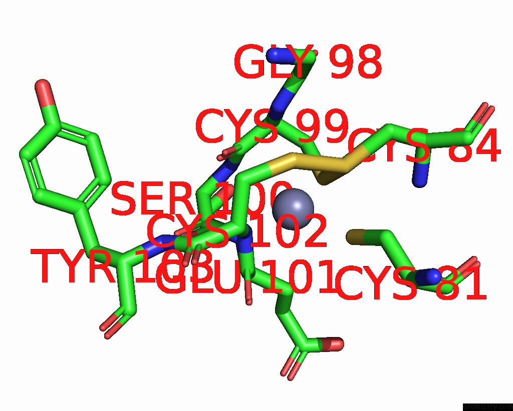
Mono view
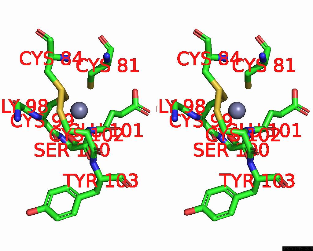
Stereo pair view
Mono view
Stereo pair view
A full contact list of Zinc with other atoms in the Zn binding
site number 1 of Crystal Structure of Mcsb Kinase Domain Complexed with Mcsa. within 5.0Å range:
|
Zinc binding site 2 out of 2 in 8wtc
Go back to
Zinc binding site 2 out
of 2 in the Crystal Structure of Mcsb Kinase Domain Complexed with Mcsa.
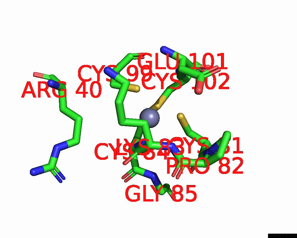
Mono view
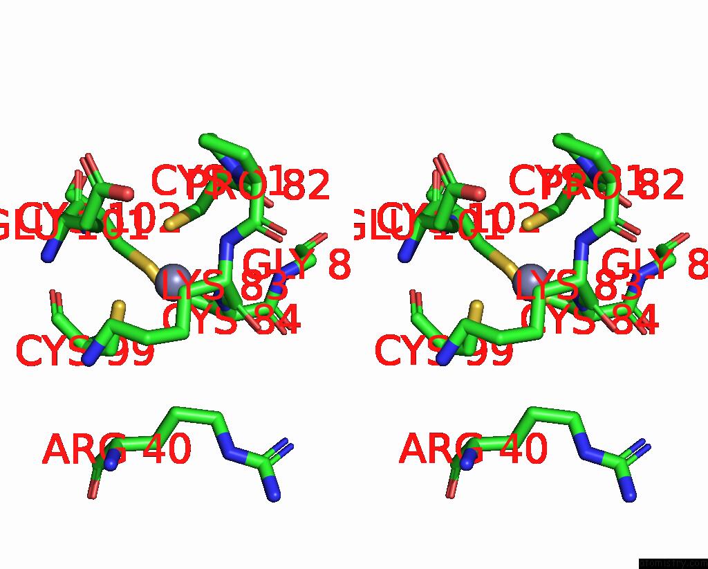
Stereo pair view
Mono view
Stereo pair view
A full contact list of Zinc with other atoms in the Zn binding
site number 2 of Crystal Structure of Mcsb Kinase Domain Complexed with Mcsa. within 5.0Å range:
|
Reference:
M.Arifuzzaman,
E.Kwon,
D.Y.Kim.
Structural Insights Into the Regulation of Protein-Arginine Kinase Mcsb By Mcsa. Proc.Natl.Acad.Sci.Usa V. 121 12121 2024.
ISSN: ESSN 1091-6490
PubMed: 38625935
DOI: 10.1073/PNAS.2320312121
Page generated: Fri Aug 22 15:20:31 2025
ISSN: ESSN 1091-6490
PubMed: 38625935
DOI: 10.1073/PNAS.2320312121
Last articles
Mn in 9LJUMn in 9LJW
Mn in 9LJS
Mn in 9LJR
Mn in 9LJT
Mn in 9LJV
Mg in 9UA2
Mg in 9R96
Mg in 9VM1
Mg in 9P01