Zinc »
PDB 7v9n-7vsp »
7vib »
Zinc in PDB 7vib: Crystal Structure of Human ACE2 and Gx/P2V Rbd
Enzymatic activity of Crystal Structure of Human ACE2 and Gx/P2V Rbd
All present enzymatic activity of Crystal Structure of Human ACE2 and Gx/P2V Rbd:
3.4.17.23;
3.4.17.23;
Protein crystallography data
The structure of Crystal Structure of Human ACE2 and Gx/P2V Rbd, PDB code: 7vib
was solved by
Y.Guo,
W.Cao,
N.Jia,
W.Wang,
S.Yuan,
Y.Wang,
with X-Ray Crystallography technique. A brief refinement statistics is given in the table below:
| Resolution Low / High (Å) | 47.29 / 3.20 |
| Space group | P 1 21 1 |
| Cell size a, b, c (Å), α, β, γ (°) | 81.672, 120.15, 107.885, 90, 96.61, 90 |
| R / Rfree (%) | 20.4 / 25.8 |
Zinc Binding Sites:
The binding sites of Zinc atom in the Crystal Structure of Human ACE2 and Gx/P2V Rbd
(pdb code 7vib). This binding sites where shown within
5.0 Angstroms radius around Zinc atom.
In total 2 binding sites of Zinc where determined in the Crystal Structure of Human ACE2 and Gx/P2V Rbd, PDB code: 7vib:
Jump to Zinc binding site number: 1; 2;
In total 2 binding sites of Zinc where determined in the Crystal Structure of Human ACE2 and Gx/P2V Rbd, PDB code: 7vib:
Jump to Zinc binding site number: 1; 2;
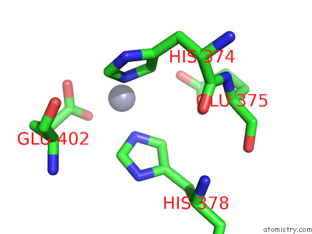
Zinc binding site 1 out of 2 in 7vib
Go back to
Zinc binding site 1 out
of 2 in the Crystal Structure of Human ACE2 and Gx/P2V Rbd

Mono view

Stereo pair view
Mono view
Stereo pair view
A full contact list of Zinc with other atoms in the Zn binding
site number 1 of Crystal Structure of Human ACE2 and Gx/P2V Rbd within 5.0Å range:
|
Zinc binding site 2 out of 2 in 7vib
Go back to
Zinc binding site 2 out
of 2 in the Crystal Structure of Human ACE2 and Gx/P2V Rbd
Mono view

Stereo pair view
Mono view
Stereo pair view
A full contact list of Zinc with other atoms in the Zn binding
site number 2 of Crystal Structure of Human ACE2 and Gx/P2V Rbd within 5.0Å range:
|
Reference:
Y.Guo,
W.Cao,
N.Jia,
W.Wang,
S.Yuan,
Y.Wang.
Crystal Structure of Human ACE2 and Gx/P2V Rbd To Be Published.
Page generated: Fri Aug 22 05:41:58 2025
Last articles
Zn in 8F5PZn in 8F5O
Zn in 8F5V
Zn in 8F4Y
Zn in 8F4S
Zn in 8F1K
Zn in 8F1C
Zn in 8F1J
Zn in 8F1D
Zn in 8F1I