Zinc »
PDB 7uzo-7v97 »
7v0f »
Zinc in PDB 7v0f: Structure of 6-Carboxy-5,6,7,8-Tetrahydropterin Synthase Paralog QUED2 From Acinetobacter Baumannii
Enzymatic activity of Structure of 6-Carboxy-5,6,7,8-Tetrahydropterin Synthase Paralog QUED2 From Acinetobacter Baumannii
All present enzymatic activity of Structure of 6-Carboxy-5,6,7,8-Tetrahydropterin Synthase Paralog QUED2 From Acinetobacter Baumannii:
4.1.2.50;
4.1.2.50;
Protein crystallography data
The structure of Structure of 6-Carboxy-5,6,7,8-Tetrahydropterin Synthase Paralog QUED2 From Acinetobacter Baumannii, PDB code: 7v0f
was solved by
M.R.Jordan,
G.Gonzalez-Gutierrez,
D.P.Giedroc,
with X-Ray Crystallography technique. A brief refinement statistics is given in the table below:
| Resolution Low / High (Å) | 42.64 / 2.35 |
| Space group | I 4 |
| Cell size a, b, c (Å), α, β, γ (°) | 84.06, 84.06, 122.42, 90, 90, 90 |
| R / Rfree (%) | 23.7 / 27.4 |
Zinc Binding Sites:
The binding sites of Zinc atom in the Structure of 6-Carboxy-5,6,7,8-Tetrahydropterin Synthase Paralog QUED2 From Acinetobacter Baumannii
(pdb code 7v0f). This binding sites where shown within
5.0 Angstroms radius around Zinc atom.
In total 4 binding sites of Zinc where determined in the Structure of 6-Carboxy-5,6,7,8-Tetrahydropterin Synthase Paralog QUED2 From Acinetobacter Baumannii, PDB code: 7v0f:
Jump to Zinc binding site number: 1; 2; 3; 4;
In total 4 binding sites of Zinc where determined in the Structure of 6-Carboxy-5,6,7,8-Tetrahydropterin Synthase Paralog QUED2 From Acinetobacter Baumannii, PDB code: 7v0f:
Jump to Zinc binding site number: 1; 2; 3; 4;
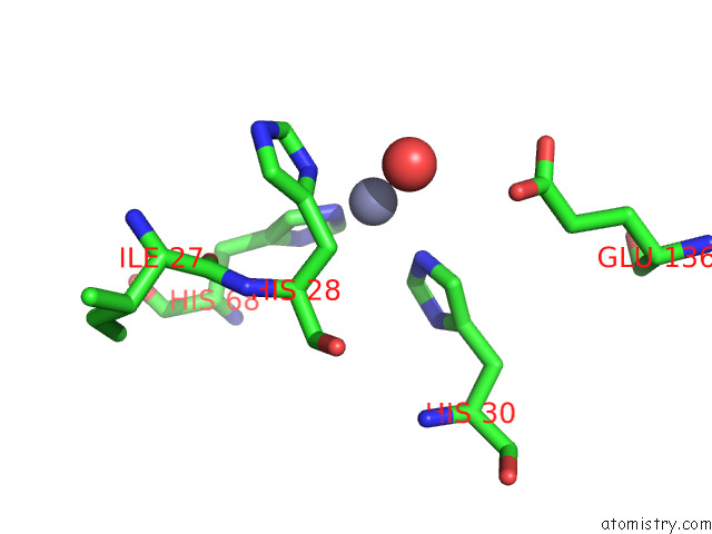
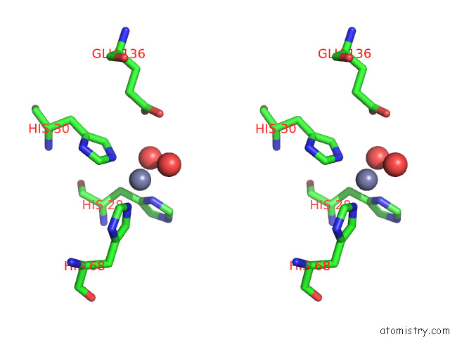
Zinc binding site 1 out of 4 in 7v0f
Go back to
Zinc binding site 1 out
of 4 in the Structure of 6-Carboxy-5,6,7,8-Tetrahydropterin Synthase Paralog QUED2 From Acinetobacter Baumannii
Mono view
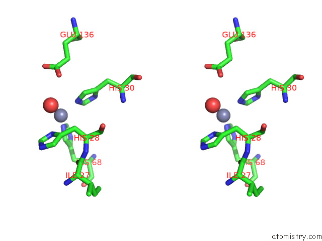
Stereo pair view
Mono view
Stereo pair view
A full contact list of Zinc with other atoms in the Zn binding
site number 1 of Structure of 6-Carboxy-5,6,7,8-Tetrahydropterin Synthase Paralog QUED2 From Acinetobacter Baumannii within 5.0Å range:
|
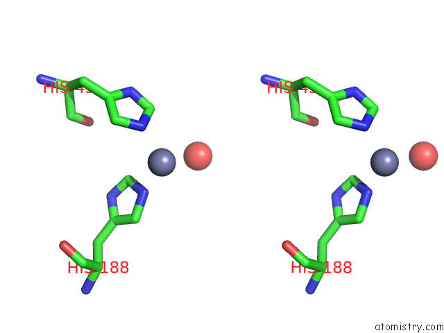
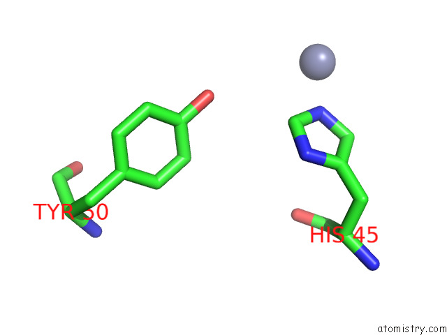
Zinc binding site 2 out of 4 in 7v0f
Go back to
Zinc binding site 2 out
of 4 in the Structure of 6-Carboxy-5,6,7,8-Tetrahydropterin Synthase Paralog QUED2 From Acinetobacter Baumannii

Mono view
Stereo pair view
Mono view
Stereo pair view
A full contact list of Zinc with other atoms in the Zn binding
site number 2 of Structure of 6-Carboxy-5,6,7,8-Tetrahydropterin Synthase Paralog QUED2 From Acinetobacter Baumannii within 5.0Å range:
|
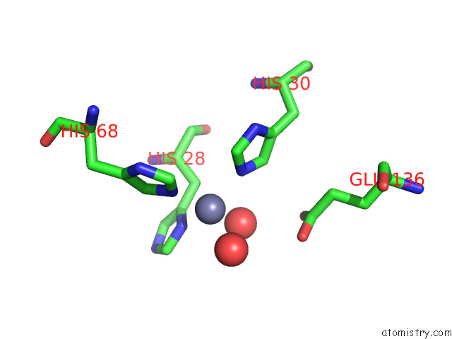
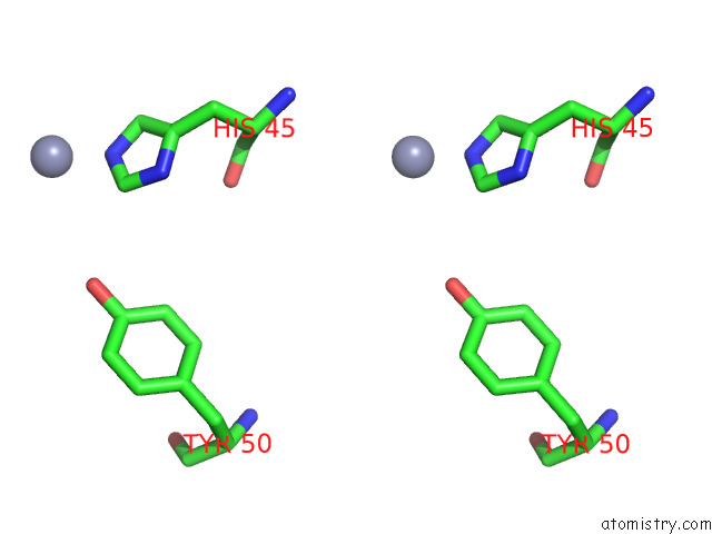
Zinc binding site 3 out of 4 in 7v0f
Go back to
Zinc binding site 3 out
of 4 in the Structure of 6-Carboxy-5,6,7,8-Tetrahydropterin Synthase Paralog QUED2 From Acinetobacter Baumannii
Mono view
Stereo pair view
Mono view
Stereo pair view
A full contact list of Zinc with other atoms in the Zn binding
site number 3 of Structure of 6-Carboxy-5,6,7,8-Tetrahydropterin Synthase Paralog QUED2 From Acinetobacter Baumannii within 5.0Å range:
|
Zinc binding site 4 out of 4 in 7v0f
Go back to
Zinc binding site 4 out
of 4 in the Structure of 6-Carboxy-5,6,7,8-Tetrahydropterin Synthase Paralog QUED2 From Acinetobacter Baumannii
Mono view
Stereo pair view
Mono view
Stereo pair view
A full contact list of Zinc with other atoms in the Zn binding
site number 4 of Structure of 6-Carboxy-5,6,7,8-Tetrahydropterin Synthase Paralog QUED2 From Acinetobacter Baumannii within 5.0Å range:
|
Reference:
M.R.Jordan,
G.Gonzalez-Gutierrez,
J.C.Trinidad,
D.P.Giedroc.
Metal Retention and Replacement in QUED2 Protect Queuosine-Trna Biosynthesis in Metal-Starved Acinetobacter Baumannii. Proc.Natl.Acad.Sci.Usa V. 119 30119 2022.
ISSN: ESSN 1091-6490
PubMed: 36442121
DOI: 10.1073/PNAS.2213630119
Page generated: Fri Aug 22 05:31:50 2025
ISSN: ESSN 1091-6490
PubMed: 36442121
DOI: 10.1073/PNAS.2213630119
Last articles
Zn in 8F0VZn in 8EZ2
Zn in 8EWI
Zn in 8EYF
Zn in 8EYL
Zn in 8EXY
Zn in 8EYE
Zn in 8EYD
Zn in 8EXT
Zn in 8EMQ