Zinc »
PDB 7ts8-7u5q »
7u2r »
Zinc in PDB 7u2r: Structure of Paenibacillus Sp. J14 APYC1
Protein crystallography data
The structure of Structure of Paenibacillus Sp. J14 APYC1, PDB code: 7u2r
was solved by
S.J.Hobbs,
T.Wein,
A.Lu,
B.R.Morehouse,
J.Schnabel,
R.Sorek,
P.J.Kranzusch,
with X-Ray Crystallography technique. A brief refinement statistics is given in the table below:
| Resolution Low / High (Å) | 38.14 / 1.85 |
| Space group | P 61 2 2 |
| Cell size a, b, c (Å), α, β, γ (°) | 102.868, 102.868, 147.619, 90, 90, 120 |
| R / Rfree (%) | 17.7 / 20 |
Zinc Binding Sites:
The binding sites of Zinc atom in the Structure of Paenibacillus Sp. J14 APYC1
(pdb code 7u2r). This binding sites where shown within
5.0 Angstroms radius around Zinc atom.
In total 2 binding sites of Zinc where determined in the Structure of Paenibacillus Sp. J14 APYC1, PDB code: 7u2r:
Jump to Zinc binding site number: 1; 2;
In total 2 binding sites of Zinc where determined in the Structure of Paenibacillus Sp. J14 APYC1, PDB code: 7u2r:
Jump to Zinc binding site number: 1; 2;
Zinc binding site 1 out of 2 in 7u2r
Go back to
Zinc binding site 1 out
of 2 in the Structure of Paenibacillus Sp. J14 APYC1
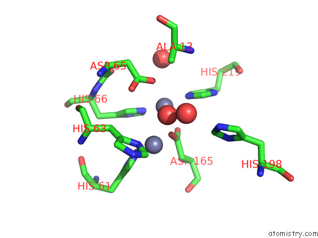
Mono view
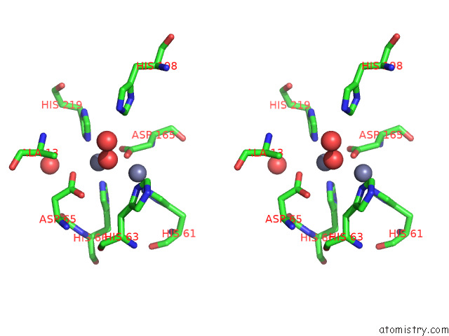
Stereo pair view
Mono view
Stereo pair view
A full contact list of Zinc with other atoms in the Zn binding
site number 1 of Structure of Paenibacillus Sp. J14 APYC1 within 5.0Å range:
|
Zinc binding site 2 out of 2 in 7u2r
Go back to
Zinc binding site 2 out
of 2 in the Structure of Paenibacillus Sp. J14 APYC1
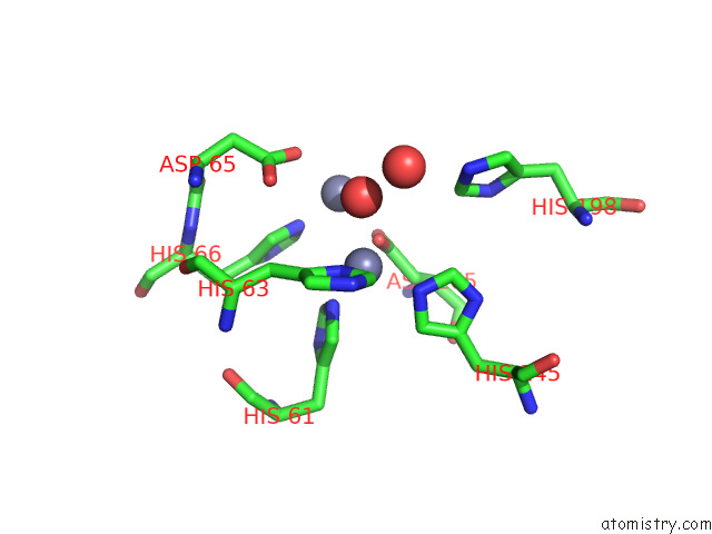
Mono view
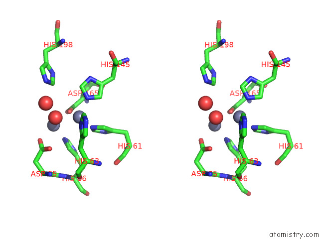
Stereo pair view
Mono view
Stereo pair view
A full contact list of Zinc with other atoms in the Zn binding
site number 2 of Structure of Paenibacillus Sp. J14 APYC1 within 5.0Å range:
|
Reference:
S.J.Hobbs,
T.Wein,
A.Lu,
B.R.Morehouse,
J.Schnabel,
A.Leavitt,
E.Yirmiya,
R.Sorek,
P.J.Kranzusch.
Phage Anti-Cbass and Anti-Pycsar Nucleases Subvert Bacterial Immunity. Nature V. 605 522 2022.
ISSN: ESSN 1476-4687
PubMed: 35395152
DOI: 10.1038/S41586-022-04716-Y
Page generated: Fri Aug 22 05:07:00 2025
ISSN: ESSN 1476-4687
PubMed: 35395152
DOI: 10.1038/S41586-022-04716-Y
Last articles
Zn in 8DPRZn in 8DPO
Zn in 8DPC
Zn in 8DJH
Zn in 8DJI
Zn in 8DI4
Zn in 8DJ9
Zn in 8DEY
Zn in 8DGT
Zn in 8DH6