Zinc »
PDB 7ts8-7u5q »
7tw7 »
Zinc in PDB 7tw7: Structure of NSP14 N7-Methyltransferase Domain Fused with Telsam Bound to Sam
Protein crystallography data
The structure of Structure of NSP14 N7-Methyltransferase Domain Fused with Telsam Bound to Sam, PDB code: 7tw7
was solved by
J.Kottur,
A.K.Aggarwal,
with X-Ray Crystallography technique. A brief refinement statistics is given in the table below:
| Resolution Low / High (Å) | 36.21 / 1.62 |
| Space group | P 65 |
| Cell size a, b, c (Å), α, β, γ (°) | 108.774, 108.774, 48.527, 90, 90, 120 |
| R / Rfree (%) | 17.9 / 21.8 |
Zinc Binding Sites:
The binding sites of Zinc atom in the Structure of NSP14 N7-Methyltransferase Domain Fused with Telsam Bound to Sam
(pdb code 7tw7). This binding sites where shown within
5.0 Angstroms radius around Zinc atom.
In total only one binding site of Zinc was determined in the Structure of NSP14 N7-Methyltransferase Domain Fused with Telsam Bound to Sam, PDB code: 7tw7:
In total only one binding site of Zinc was determined in the Structure of NSP14 N7-Methyltransferase Domain Fused with Telsam Bound to Sam, PDB code: 7tw7:
Zinc binding site 1 out of 1 in 7tw7
Go back to
Zinc binding site 1 out
of 1 in the Structure of NSP14 N7-Methyltransferase Domain Fused with Telsam Bound to Sam
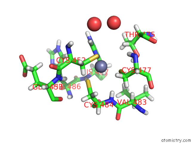
Mono view
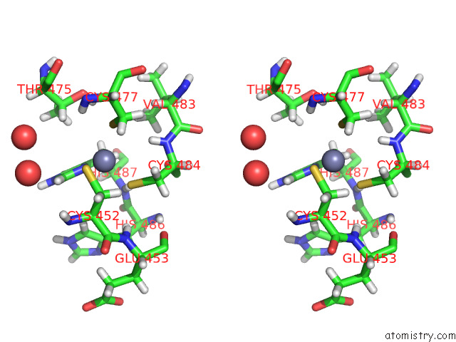
Stereo pair view
Mono view
Stereo pair view
A full contact list of Zinc with other atoms in the Zn binding
site number 1 of Structure of NSP14 N7-Methyltransferase Domain Fused with Telsam Bound to Sam within 5.0Å range:
|
Reference:
J.Kottur,
O.Rechkoblit,
R.Quintana-Feliciano,
D.Sciaky,
A.K.Aggarwal.
High-Resolution Structures of the Sars-Cov-2 N7-Methyltransferase Inform Therapeutic Development. Nat.Struct.Mol.Biol. V. 29 850 2022.
ISSN: ESSN 1545-9985
PubMed: 36075969
DOI: 10.1038/S41594-022-00828-1
Page generated: Fri Aug 22 05:05:06 2025
ISSN: ESSN 1545-9985
PubMed: 36075969
DOI: 10.1038/S41594-022-00828-1
Last articles
Zn in 8PGUZn in 8PGS
Zn in 8PGR
Zn in 8PGQ
Zn in 8PGO
Zn in 8PGP
Zn in 8PGN
Zn in 8PGL
Zn in 8PGJ
Zn in 8PGK