Zinc »
PDB 7ml7-7mot »
7mmb »
Zinc in PDB 7mmb: Crystal Structure of Hcv NS3/4A D168A Protease in Complex with NR01- 127
Protein crystallography data
The structure of Crystal Structure of Hcv NS3/4A D168A Protease in Complex with NR01- 127, PDB code: 7mmb
was solved by
J.Zephyr,
C.A.Schiffer,
with X-Ray Crystallography technique. A brief refinement statistics is given in the table below:
| Resolution Low / High (Å) | 23.84 / 1.99 |
| Space group | P 21 21 21 |
| Cell size a, b, c (Å), α, β, γ (°) | 54.316, 59.492, 58.731, 90, 90, 90 |
| R / Rfree (%) | 18.3 / 22.3 |
Zinc Binding Sites:
The binding sites of Zinc atom in the Crystal Structure of Hcv NS3/4A D168A Protease in Complex with NR01- 127
(pdb code 7mmb). This binding sites where shown within
5.0 Angstroms radius around Zinc atom.
In total only one binding site of Zinc was determined in the Crystal Structure of Hcv NS3/4A D168A Protease in Complex with NR01- 127, PDB code: 7mmb:
In total only one binding site of Zinc was determined in the Crystal Structure of Hcv NS3/4A D168A Protease in Complex with NR01- 127, PDB code: 7mmb:
Zinc binding site 1 out of 1 in 7mmb
Go back to
Zinc binding site 1 out
of 1 in the Crystal Structure of Hcv NS3/4A D168A Protease in Complex with NR01- 127
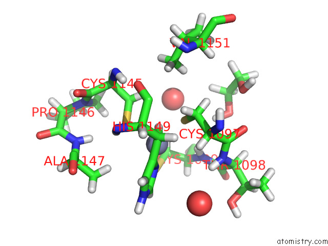
Mono view
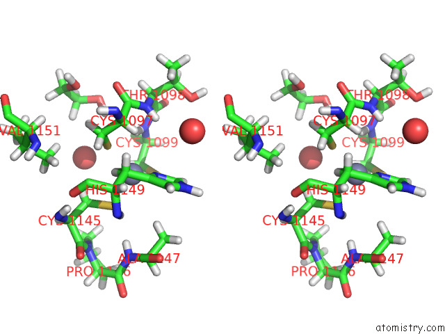
Stereo pair view
Mono view
Stereo pair view
A full contact list of Zinc with other atoms in the Zn binding
site number 1 of Crystal Structure of Hcv NS3/4A D168A Protease in Complex with NR01- 127 within 5.0Å range:
|
Reference:
J.Zephyr,
D.Nageswara Rao,
S.V.Vo,
M.Henes,
K.Kosovrasti,
A.N.Matthew,
A.K.Hedger,
J.Timm,
E.T.Chan,
A.Ali,
N.Kurt Yilmaz,
C.A.Schiffer.
Deciphering the Molecular Mechanism of Hcv Protease Inhibitor Fluorination As A General Approach to Avoid Drug Resistance. J.Mol.Biol. V. 434 67503 2022.
ISSN: ESSN 1089-8638
PubMed: 35183560
DOI: 10.1016/J.JMB.2022.167503
Page generated: Fri Aug 22 02:21:24 2025
ISSN: ESSN 1089-8638
PubMed: 35183560
DOI: 10.1016/J.JMB.2022.167503
Last articles
Zn in 8CJ7Zn in 8CHX
Zn in 8CDB
Zn in 8CEF
Zn in 8CGW
Zn in 8CGV
Zn in 8CGP
Zn in 8CGK
Zn in 8CGD
Zn in 8CG9